• 文献检索
  • 文档翻译
  • 深度研究
  • 学术资讯
  • Suppr Zotero 插件Zotero 插件
  • 邀请有礼
  • 套餐&价格
  • 历史记录
应用&插件
Suppr Zotero 插件Zotero 插件浏览器插件Mac 客户端Windows 客户端微信小程序
定价
高级版会员购买积分包购买API积分包
服务
文献检索文档翻译深度研究API 文档MCP 服务
关于我们
关于 Suppr公司介绍联系我们用户协议隐私条款
关注我们

Suppr 超能文献

核心技术专利:CN118964589B侵权必究
粤ICP备2023148730 号-1Suppr @ 2026

文献检索

告别复杂PubMed语法,用中文像聊天一样搜索,搜遍4000万医学文献。AI智能推荐,让科研检索更轻松。

立即免费搜索

文件翻译

保留排版,准确专业,支持PDF/Word/PPT等文件格式,支持 12+语言互译。

免费翻译文档

深度研究

AI帮你快速写综述,25分钟生成高质量综述,智能提取关键信息,辅助科研写作。

立即免费体验

家族性多灶性微结节性肺细胞增生症伴 TSC1 新剪接突变:一家三例。

Familial multifocal micronodular pneumocyte hyperplasia with a novel splicing mutation in TSC1: Three cases in one family.

机构信息

Department of Respiratory Medicine, Faculty of Medicine and Graduate School of Medicine, Hokkaido University, Sapporo, Hokkaido, Japan.

Center for Clinical Genomics, Kanazawa Medical University Hospital, Ishikawa, Japan.

出版信息

PLoS One. 2019 Feb 22;14(2):e0212370. doi: 10.1371/journal.pone.0212370. eCollection 2019.

DOI:10.1371/journal.pone.0212370
PMID:30794603
原文链接:https://pmc.ncbi.nlm.nih.gov/articles/PMC6386448/
Abstract

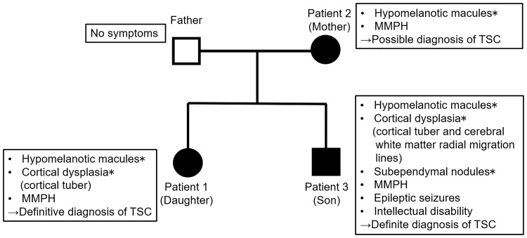
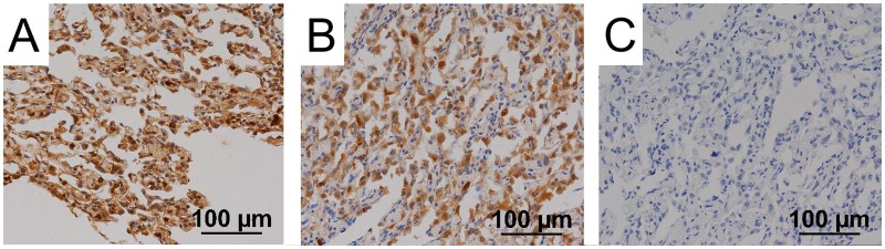
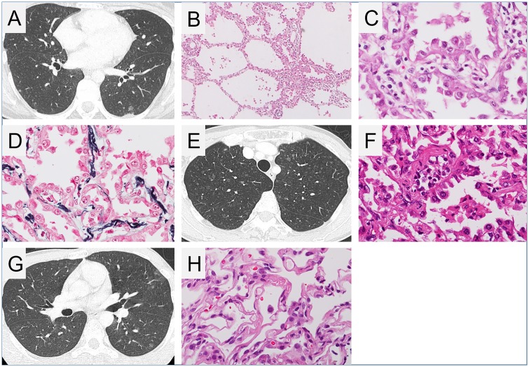
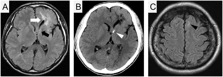
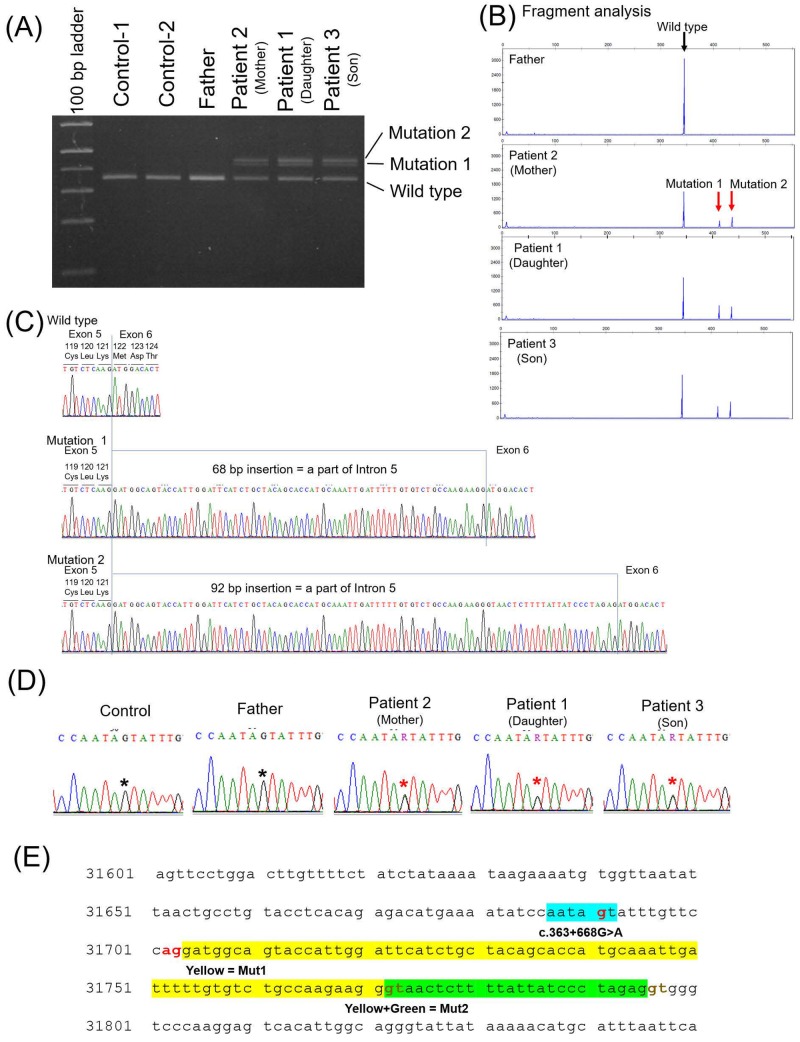
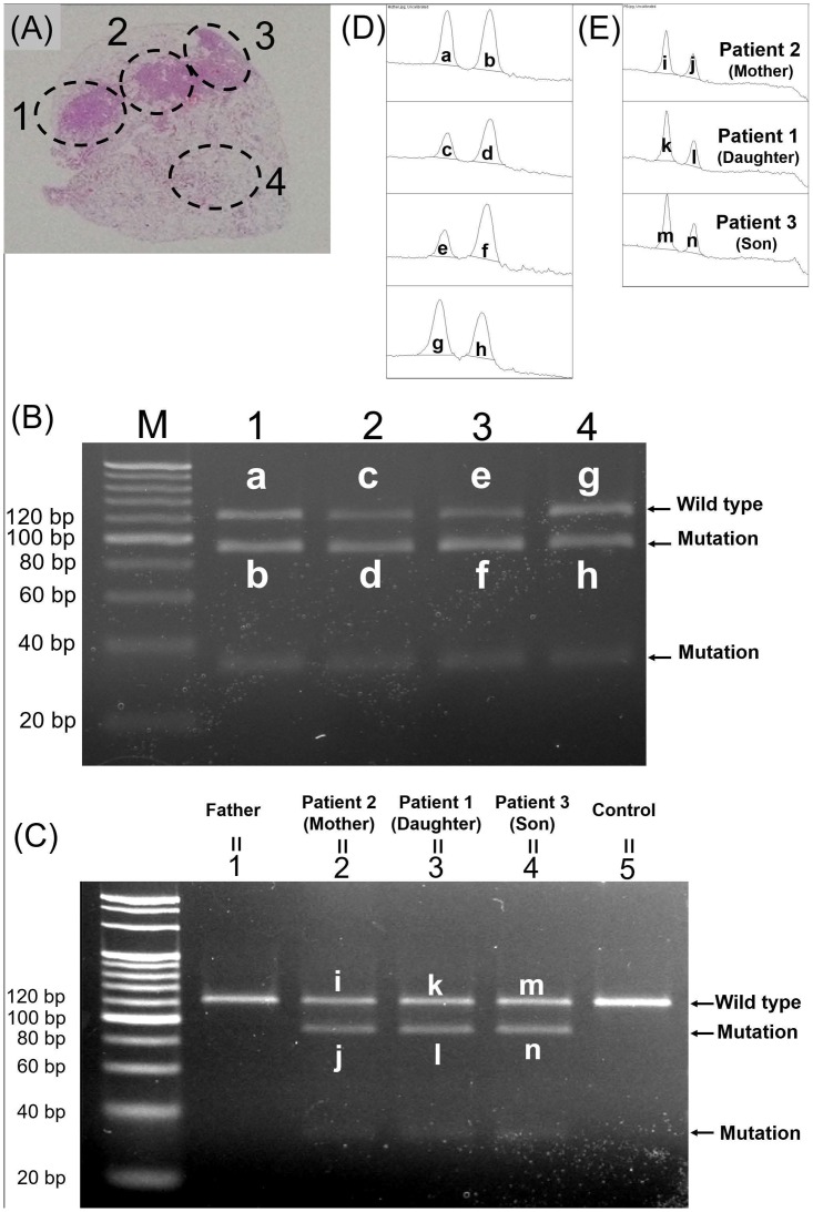
Multifocal micronodular pneumocyte hyperplasia (MMPH) is a rare pulmonary disease, generally manifesting as a tuberous sclerosis complex (TSC), characterised by multiple, small ground-glass nodular shadows on chest computed tomography (CT). Histological examination typically reveals multicentric, well-demarcated, nodular type II pneumocystic growth. Herein, we describe three cases of this rare pulmonary disease occurring within one family. Using reverse transcription polymerase chain reaction (RT-PCR) and direct DNA sequencing, we identified a novel germline mutation, a point mutation in TSC1 intron 5, which yielded a splice variant and loss of function of TSC1. Furthermore, immunohistochemical staining indicated the expression of phospho-p70S6K and phospho-4E-BP1, suggesting that TSC1 function was impaired by the novel gene mutation in MMPH cells.

摘要

多灶性微结节性肺泡细胞增生症(MMPH)是一种罕见的肺部疾病,通常表现为结节性硬化症复合征(TSC),其特征是胸部计算机断层扫描(CT)上存在多个、小的磨玻璃结节状阴影。组织学检查通常显示多中心、边界清楚的结节型 II 型肺泡细胞增生。本文描述了一家族中发生的三例这种罕见的肺部疾病。通过逆转录聚合酶链反应(RT-PCR)和直接 DNA 测序,我们鉴定出一种新型胚系突变,即 TSC1 内含子 5 中的点突变,导致剪接变异体和 TSC1 功能丧失。此外,免疫组织化学染色表明磷酸化-p70S6K 和磷酸化-4E-BP1 的表达,提示 MMPH 细胞中的新型基因突变导致 TSC1 功能受损。

https://cdn.ncbi.nlm.nih.gov/pmc/blobs/24a4/6386448/fce68faa6c31/pone.0212370.g006.jpg
https://cdn.ncbi.nlm.nih.gov/pmc/blobs/24a4/6386448/ce48e17a737f/pone.0212370.g001.jpg
https://cdn.ncbi.nlm.nih.gov/pmc/blobs/24a4/6386448/b5951f8e9a76/pone.0212370.g002.jpg
https://cdn.ncbi.nlm.nih.gov/pmc/blobs/24a4/6386448/d094be68cc27/pone.0212370.g003.jpg
https://cdn.ncbi.nlm.nih.gov/pmc/blobs/24a4/6386448/da75ee723cda/pone.0212370.g004.jpg
https://cdn.ncbi.nlm.nih.gov/pmc/blobs/24a4/6386448/ebcf151bab90/pone.0212370.g005.jpg
https://cdn.ncbi.nlm.nih.gov/pmc/blobs/24a4/6386448/fce68faa6c31/pone.0212370.g006.jpg

相似文献

1
Familial multifocal micronodular pneumocyte hyperplasia with a novel splicing mutation in TSC1: Three cases in one family.家族性多灶性微结节性肺细胞增生症伴 TSC1 新剪接突变:一家三例。
PLoS One. 2019 Feb 22;14(2):e0212370. doi: 10.1371/journal.pone.0212370. eCollection 2019.
2
Loss of heterozygosity on tuberous sclerosis complex genes in multifocal micronodular pneumocyte hyperplasia.结节性硬化症复合物基因杂合性缺失在多灶性微结节性肺泡细胞增生症中的研究。
Mod Pathol. 2010 Sep;23(9):1251-60. doi: 10.1038/modpathol.2010.114. Epub 2010 Jun 4.
3
Two Cases of Tuberous Sclerosis Complex Suggestive of Complicating Multifocal Micronodular Pneumocyte Hyperplasia: A Case Report.两例结节性硬化症合并多灶性微小结节性肺细胞增生症:病例报告
J UOEH. 2017;39(2):133-141. doi: 10.7888/juoeh.39.133.
4
Novel genetic characteristics of multifocal micronodular pneumocyte hyperplasia (MMPH): a case report with frequent BRAF mutations analyzed by next-generation sequencing supporting benign behaviors of MMPH.多灶性微结节性肺泡细胞增生症(MMPH)的新遗传特征:一例 BRAF 突变频繁的病例报告,通过下一代测序支持 MMPH 的良性行为。
Virchows Arch. 2021 Sep;479(3):637-641. doi: 10.1007/s00428-020-03013-1. Epub 2021 Jan 14.
5
Multifocal micronodular pneumocyte hyperplasia lacking typical clinical features of the tuberous sclerosis complex: a case report and literature review.多灶性微结节性肺泡细胞增生症,缺乏结节性硬化症的典型临床特征:病例报告及文献复习。
BMC Pulm Med. 2022 Mar 5;22(1):77. doi: 10.1186/s12890-022-01849-8.
6
[A case of multifocal micronodular pneumocyte hyperplasia without clinical findings of tuberous sclerosis].[一例无结节性硬化症临床症状的多灶性微小结节性肺细胞增生症]
Nihon Kokyuki Gakkai Zasshi. 2011 May;49(5):355-9.
7
Pathogenesis of multifocal micronodular pneumocyte hyperplasia and lymphangioleiomyomatosis in tuberous sclerosis and association with tuberous sclerosis genes TSC1 and TSC2.结节性硬化症中多灶性微小结节性肺细胞增生症和淋巴管平滑肌瘤病的发病机制及其与结节性硬化症基因TSC1和TSC2的关联
Pathol Int. 2001 Aug;51(8):585-94. doi: 10.1046/j.1440-1827.2001.01242.x.
8
Multifocal micronodular pneumocyte hyperplasia and lymphangioleiomyomatosis in tuberous sclerosis with a TSC2 gene.伴有TSC2基因的结节性硬化症中的多灶性微小结节性肺细胞增生和淋巴管平滑肌瘤病。
Mod Pathol. 2001 Jun;14(6):609-14. doi: 10.1038/modpathol.3880359.
9
Clinicopathological analysis of multifocal micronodular pneumocyte hyperplasia associated with tuberous sclerosis in Japan.日本结节性硬化症相关多灶性微小结节性肺细胞增生的临床病理分析
Respirology. 2008 Nov;13(7):1076-81. doi: 10.1111/j.1440-1843.2008.01382.x. Epub 2008 Aug 10.
10
Multifocal micronodular pneumocyte hyperplasia associated with tuberous sclerosis.与结节性硬化症相关的多灶性微小结节性肺细胞增生症
Acta Radiol. 2005 Feb;46(1):37-40. doi: 10.1080/02841850510020860.

引用本文的文献

1
Genetic analysis of a novel TSC1 splice mutation causing tuberous sclerosis without neurological phenotypes.导致无神经学表型的结节性硬化症的新型TSC1剪接突变的基因分析。
Sci Rep. 2025 Jul 9;15(1):24780. doi: 10.1038/s41598-025-07906-6.
2
[Is It Possible to Diagnose Lymphangioleiomyomatosis or Multifocal Micronodular Pneumocyte Hyperplasia Without Visible Lesions on the Chest Tomography?].[胸部断层扫描无可见病变时能否诊断淋巴管平滑肌瘤病或多灶性微小结节性肺细胞增生?]
Open Respir Arch. 2021 Jul 26;3(3):100120. doi: 10.1016/j.opresp.2021.100120. eCollection 2021 Jul-Sep.
3
Genotype and Phenotype Landscape of 283 Japanese Patients with Tuberous Sclerosis Complex.

本文引用的文献

1
Clinical Course of Histologically Proven Multifocal Micronodular Pneumocyte Hyperplasia in Tuberous Sclerosis Complex: A Case Series and Comparison with Lymphangiomyomatosis.组织学证实的结节性肺细胞增生症在结节性硬化症中的临床过程:病例系列及与淋巴管平滑肌瘤病的比较。
Respiration. 2018;95(5):310-316. doi: 10.1159/000486101. Epub 2018 Jan 24.
2
The genomic landscape of tuberous sclerosis complex.结节性硬化症的基因组景观。
Nat Commun. 2017 Jun 15;8:15816. doi: 10.1038/ncomms15816.
3
Whole Exome Sequencing Identifies TSC1/TSC2 Biallelic Loss as the Primary and Sufficient Driver Event for Renal Angiomyolipoma Development.
283 例日本结节性硬化症患者的基因型和表型全景分析。
Int J Mol Sci. 2022 Sep 22;23(19):11175. doi: 10.3390/ijms231911175.
4
Prevalence, parameters, and pathogenic mechanisms for splice-altering acceptor variants that disrupt the AG exclusion zone.破坏AG排除区的剪接改变受体变体的患病率、参数和致病机制。
HGG Adv. 2022 Jun 25;3(4):100125. doi: 10.1016/j.xhgg.2022.100125. eCollection 2022 Oct 13.
5
Multifocal micronodular pneumocyte hyperplasia with a novel mutation in TSC1: a case report.多灶性微结节性肺泡细胞增生症伴 TSC1 新突变:病例报告。
Croat Med J. 2021 Oct 31;62(5):523-527. doi: 10.3325/cmj.2021.62.523.
6
Resolution of multifocal micronodular pneumocyte hyperplasia with everolimus in a patient with tuberous sclerosis complex.依维莫司治疗结节性硬化症患者的多灶性微小结节性肺细胞增生症的疗效
Respir Med Case Rep. 2021 Oct 6;34:101526. doi: 10.1016/j.rmcr.2021.101526. eCollection 2021.
全外显子组测序确定TSC1/TSC2双等位基因缺失是肾血管平滑肌脂肪瘤发生的主要且充分的驱动事件。
PLoS Genet. 2016 Aug 5;12(8):e1006242. doi: 10.1371/journal.pgen.1006242. eCollection 2016 Aug.
4
Mosaic and Intronic Mutations in TSC1/TSC2 Explain the Majority of TSC Patients with No Mutation Identified by Conventional Testing.TSC1/TSC2中的镶嵌突变和内含子突变解释了大多数经传统检测未发现突变的结节性硬化症患者的病因。
PLoS Genet. 2015 Nov 5;11(11):e1005637. doi: 10.1371/journal.pgen.1005637. eCollection 2015 Nov.
5
Tuberous Sclerosis Complex: State-of-the-Art Review with a Focus on Pulmonary Involvement.结节性硬化症:聚焦肺部受累的最新综述
Lung. 2015 Oct;193(5):619-27. doi: 10.1007/s00408-015-9750-6. Epub 2015 Jun 24.
6
Tuberous sclerosis complex surveillance and management: recommendations of the 2012 International Tuberous Sclerosis Complex Consensus Conference.结节性硬化症的监测和管理:2012 年国际结节性硬化症共识会议的建议。
Pediatr Neurol. 2013 Oct;49(4):255-65. doi: 10.1016/j.pediatrneurol.2013.08.002.
7
Tuberous sclerosis complex diagnostic criteria update: recommendations of the 2012 Iinternational Tuberous Sclerosis Complex Consensus Conference.结节性硬化症综合诊断标准更新:2012 年国际结节性硬化症共识会议推荐。
Pediatr Neurol. 2013 Oct;49(4):243-54. doi: 10.1016/j.pediatrneurol.2013.08.001.
8
Trends in the prevalence of tuberous sclerosis complex manifestations: an epidemiological study of 166 Japanese patients.结节性硬化症表现的流行趋势:166 例日本患者的流行病学研究。
PLoS One. 2013 May 17;8(5):e63910. doi: 10.1371/journal.pone.0063910. Print 2013.
9
Mutational analysis of TSC1 and TSC2 in Japanese patients with tuberous sclerosis complex revealed higher incidence of TSC1 patients than previously reported.对日本结节性硬化症患者的 TSC1 和 TSC2 进行突变分析显示,TSC1 患者的发病率高于以往报道。
J Hum Genet. 2013 Apr;58(4):216-25. doi: 10.1038/jhg.2013.3. Epub 2013 Feb 7.
10
Applying and testing the conveniently optimized enzyme mismatch cleavage method to clinical DNA diagnosis.应用和测试经过优化的方便酶错配切割法进行临床 DNA 诊断。
Mol Genet Metab. 2012 Nov;107(3):580-5. doi: 10.1016/j.ymgme.2012.09.008. Epub 2012 Sep 10.