Jiang Meng-Nan, Zhou Yu-Yang, Hua Di-Hao, Yang Jia-Yi, Hu Man-Li, Xing Yi-Qiao
Eye Center, Renmin Hospital of Wuhan University, Wuhan, China.
College of Veterinary Medicine, Huazhong Agricultural University, Wuhan, China.
Front Neurosci. 2019 Feb 11;13:87. doi: 10.3389/fnins.2019.00087. eCollection 2019.
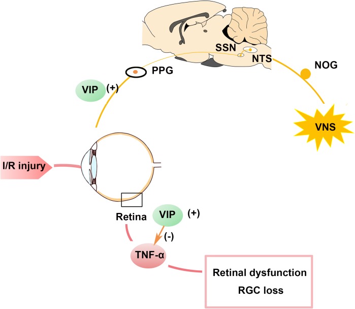
The present study aimed to investigate whether cervical vagal nerve stimulation (VNS) could prevent retinal ganglion cell (RGC) loss and retinal dysfunction after ischemia/reperfusion (I/R) injury. First, rats were randomly divided into sham group ( = 4) and VNS group ( = 12). Activation of the nodose ganglia (NOG), nucleus of the solitary tract (NTS), superior salivatory nucleus (SSN), and pterygopalatine ganglion (PPG) neural circuit were evaluated by c-fos expression at 0 h after sham VNS and at 0 h ( = 4), 6 h ( = 4), 72 h ( = 4) after VNS. Secondly, rats were randomly assigned to I/R group (pressure-induced retinal ischemia for 1 h and reperfusion for 1 h in the right eye, = 16) and I/R+VNS group (right cervical VNS for 2 h during the I/R period, = 16). The left eye of each rat served as a control. Electroretinogram (ERG), RGC numbers, tumor necrosis factor-α (TNF-α) and vasoactive intestinal polypeptide (VIP) levels in retina were determined. Additionally, the level of VIP in PPG was evaluated. In the first part of the study, compared with the sham group, the VNS group exhibited significantly increased expression of c-fos in NOG, NTS, SSN, and PPG tissues at 0, 6, and 72 h. In the second part of the study, compared with left eyes, retinal function in right eyes (as assessed by the a-wave, b-wave and the oscillatory potential amplitudes of ERG and RGC data) was significantly decreased by I/R. The decreased retinal function was attenuated by VNS. In addition, I/R induced an increase in inflammation, which was reflected by elevated TNF-α expression in the retina. VNS significantly attenuated the increase in I/R-induced inflammation. Moreover, VIP expression in the retina and PPG, which may contribute to the inhibition of the inflammatory response, was significantly increased after VNS. VNS could protect against retinal I/R injury by downregulating TNF-α. Upregulation of VIP expression due to activation of the NOG-NTS-SSN-PPG neural circuit may underlie to the protective effects of VNS.
本研究旨在探讨颈迷走神经刺激(VNS)是否能预防缺血/再灌注(I/R)损伤后视网膜神经节细胞(RGC)丢失和视网膜功能障碍。首先,将大鼠随机分为假手术组(n = 4)和VNS组(n = 12)。通过假手术VNS后0小时以及VNS后0小时(n = 4)、6小时(n = 4)、72小时(n = 4)时的c-fos表达来评估结状神经节(NOG)、孤束核(NTS)、上涎核(SSN)和翼腭神经节(PPG)神经回路的激活情况。其次,将大鼠随机分为I/R组(右眼进行压力诱导的视网膜缺血1小时和再灌注1小时,n = 16)和I/R + VNS组(在I/R期间右侧颈迷走神经刺激2小时,n = 16)。每只大鼠的左眼作为对照。测定视网膜电图(ERG)、RGC数量、肿瘤坏死因子-α(TNF-α)和血管活性肠肽(VIP)水平。此外,评估PPG中VIP的水平。在研究的第一部分,与假手术组相比,VNS组在0、6和72小时时NOG、NTS、SSN和PPG组织中的c-fos表达显著增加。在研究的第二部分,与左眼相比,I/R使右眼的视网膜功能(通过ERG的a波、b波和振荡电位振幅以及RGC数据评估)显著降低。VNS减轻了视网膜功能的下降。此外,I/R诱导炎症增加,这通过视网膜中TNF-α表达升高得以体现。VNS显著减轻了I/R诱导的炎症增加。而且,VNS后视网膜和PPG中可能有助于抑制炎症反应的VIP表达显著增加。VNS可通过下调TNF-α来保护视网膜免受I/R损伤。由于NOG-NTS-SSN-PPG神经回路激活导致的VIP表达上调可能是VNS保护作用的基础。