Huang Yi, Chen Jui-Cheng, Chen Chun-Ming, Tsai Chon-Haw, Lu Ming-Kuei
Graduate Institute of Biomedical Sciences, Medical College, China Medical University, Taichung, Taiwan.
Neuroscience Laboratory, Department of Neurology, China Medical University Hospital, Taichung, Taiwan.
Front Hum Neurosci. 2019 Feb 12;13:49. doi: 10.3389/fnhum.2019.00049. eCollection 2019.
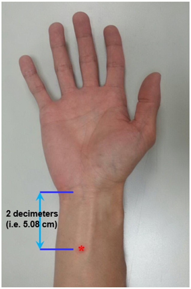
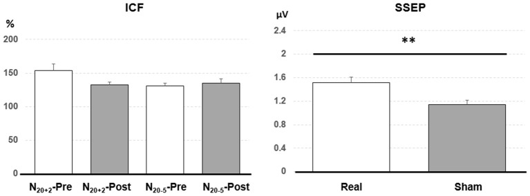
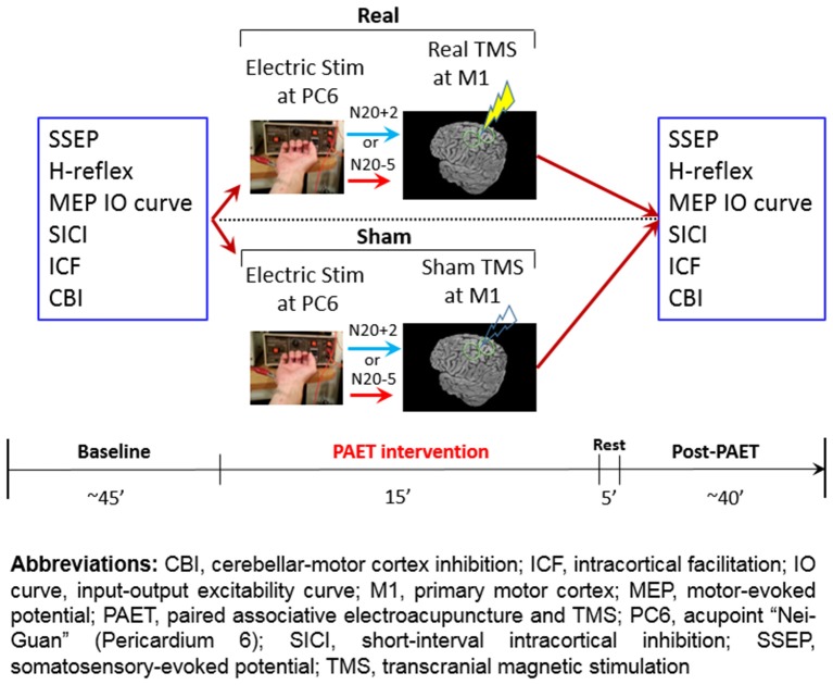
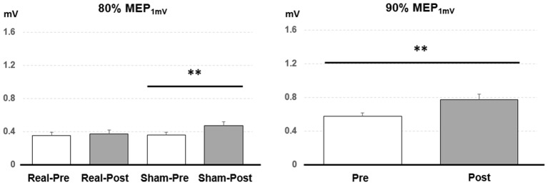
Pairing transcutaneous electric nerve stimulation (TENS) and transcranial magnetic stimulation (TMS) with specific stimulus-intervals induces associative motor plasticity at the primary motor cortex (M1). Electroacupuncture (EA) is an established medical technique in the eastern countries. This study investigates whether EA paired with TMS induces distinct M1 motor plasticity. Fifteen healthy, right-handed subjects (aged 23.6 ± 2.0 years, eight women) were studied. Two-hundred and twenty-five pairs of TMS of the left M1 preceded by right EA at acupoint "Neiguan" [Pericardium 6 (PC6), located 2 decimeters proximal from the wrist wrinkle] were respectively applied with the interstimulus interval (ISI) of individual somatosensory evoked potential (SSEP) N20 latency plus 2 ms (N20+2) and minus 5 ms (N20-5) with at least 1-week interval. The paired stimulation was delivered at a rate of 0.25 Hz. Sham TMS with a sham coil was adopted to examine the low-frequency EA influence on M1 in eleven subjects. M1 excitability was assessed by motor-evoked potential (MEP) recruitment curve with five TMS intensity levels, short-interval intracortical inhibition (SICI), intracortical facilitation (ICF) and cerebellar inhibition (CBI) at the abductor pollicis brevis (APB) muscle of the right hand before and after the EA-M1 paired associative stimulation (PAS). In addition, median nerve SSEPs and H-reflex were respectively measured to monitor somatosensory and spinal excitability. The MEP showed significantly facilitated after the sham EA-M1 PAS while tested with 80% of the TMS intensity producing on average 1 mV amplitude (i.e., MEP) in the resting APB muscle. It was also facilitated while tested with 90% MEP irrespective of the stimulation conditions. The SSEP showed a higher amplitude from the real EA-M1 PAS compared to that from the sham EA-M1 PAS. No significant change was found on SICI, ICF, CBI and H-reflex. Findings suggest that repetitive low frequency EA paired with real TMS did not induce spike-timing dependent motor plasticity but EA paired with sham TMS induced specific M1 excitability change. Complex sensory afferents with dispersed time locked to the sensorimotor cortical area could hamper instead of enhancing the induction of the spike-timing dependent plasticity (STDP) in M1.
将经皮电神经刺激(TENS)和经颅磁刺激(TMS)以特定的刺激间隔配对,可在初级运动皮层(M1)诱导联合运动可塑性。电针(EA)在东方国家是一种成熟的医学技术。本研究调查EA与TMS配对是否会诱导不同的M1运动可塑性。研究了15名健康的右利手受试者(年龄23.6±2.0岁,8名女性)。在穴位“内关”[心包经6(PC6),位于腕横纹近端2寸处]进行右侧EA后,对左侧M1进行225对TMS,分别采用个体体感诱发电位(SSEP)N20潜伏期加2毫秒(N20+2)和减5毫秒(N20-5)的刺激间隔(ISI),间隔至少1周。配对刺激以0.25赫兹的频率进行。采用带有假线圈的假TMS检查11名受试者中低频EA对M1的影响。在EA-M1配对联想刺激(PAS)前后,通过运动诱发电位(MEP)募集曲线、短间隔皮质内抑制(SICI)皮质内易化(ICF)和右手拇短展肌(APB)的小脑抑制(CBI),以五个TMS强度水平评估M1兴奋性。此外,分别测量正中神经SSEP和H反射,以监测体感和脊髓兴奋性。在静息APB肌肉中,当用平均产生1毫伏振幅(即MEP)的80%TMS强度进行测试时,假EA-M1 PAS后MEP显示出显著易化。无论刺激条件如何,用90%MEP进行测试时也出现易化。与假EA-M1 PAS相比,真实EA-M1 PAS的SSEP振幅更高。SICI、ICF、CBI和H反射未发现显著变化。研究结果表明,重复低频EA与真实TMS配对不会诱导依赖于峰时的运动可塑性,但EA与假TMS配对会诱导特定的M1兴奋性变化。与感觉运动皮层区域时间锁定分散的复杂感觉传入可能会阻碍而不是增强M1中依赖于峰时的可塑性(STDP)的诱导。