Sudjarwo Sri Agus, Eraiko Koerniasari, Sudjarwo Giftania Wardani
Department of Pharmacology, Faculty of Veterinary Medicine, Airlangga University, Surabaya, Indonesia.
Department of Conservative Dentistry, Faculty of Dentistry, Airlangga University, Surabaya, Indonesia.
J Adv Pharm Technol Res. 2019 Jan-Mar;10(1):27-32. doi: 10.4103/japtr.JAPTR_306_18.
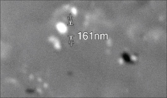
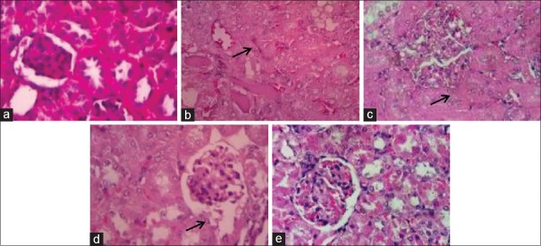
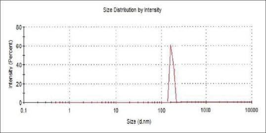
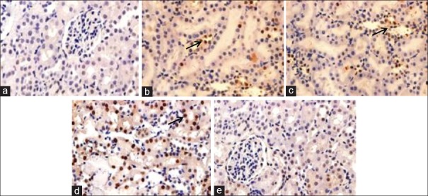
The current study was carried out to evaluate the antioxidant and anti-caspase 3 activity of chitosan- nanoparticle in against lead acetate-induced nephrotoxicity in rats. chitosan- nanoparticle was characterized by dynamic light scattering (DLS) and scanning electron microscope (SEM). The male rats were divided into control group (rats were given with distilled water), lead acetate group (rats were injected with lead acetate 15 mg/kg BW i. p), and the treatment group (rats were given the chitosan- nanoparticle 150 mg, 300 mg, 600 mg/kg BW orally and were injected with lead acetate 15 mg/kg BW). The rats blood samples were measured levels of blood urea nitrogen (BUN) and creatinine. The kidney tissues were collected to evaluate the malondialdehyde (MDA), superoxide dismutase (SOD), and glutathione peroxidase (GPx). Histological to evaluate renal damage, and immunohistochemical to analyze the expression of caspase 3. The results showed that DLS showed the size of chitosan- nanoparticle was 165.9 ± 24.18 nm. SEM images of the chitosan- nanoparticles showed an irregular shape and its the rough surface. Administration of lead acetate resulted in a significant increase in levels of the BUN, creatinine, MDA level, caspase 3 expression, and a decrease in SOD and GPx were compared with the control group. Treatment with the chitosan- nanoparticle 600 mg/kg BW significantly decreased the elevated BUN, creatinine, MDA levels, caspase 3 expression and also increase in SOD and GPx as compared to lead acetate group. The lead acetate induced loss of the normal structure of renal cells and necrosis, whereas treated with chitosan- nanoparticle improved renal cell necrosis. This study indicates that chitosan- nanoparticles appeared to be a promising agent for protection against lead-induced nephrotoxicity through increasing antioxidant and inhibiting caspase 3 expression.
本研究旨在评估壳聚糖纳米颗粒对醋酸铅诱导的大鼠肾毒性的抗氧化和抗半胱天冬酶3活性。通过动态光散射(DLS)和扫描电子显微镜(SEM)对壳聚糖纳米颗粒进行表征。将雄性大鼠分为对照组(给予蒸馏水)、醋酸铅组(腹腔注射15 mg/kg体重的醋酸铅)和治疗组(口服150 mg、300 mg、600 mg/kg体重的壳聚糖纳米颗粒并腹腔注射15 mg/kg体重的醋酸铅)。检测大鼠血液样本中的血尿素氮(BUN)和肌酐水平。收集肾脏组织评估丙二醛(MDA)、超氧化物歧化酶(SOD)和谷胱甘肽过氧化物酶(GPx)。进行组织学检查以评估肾脏损伤,并进行免疫组织化学分析以分析半胱天冬酶3的表达。结果显示,DLS表明壳聚糖纳米颗粒的大小为165.9±24.18 nm。壳聚糖纳米颗粒的SEM图像显示其形状不规则且表面粗糙。与对照组相比,给予醋酸铅导致BUN、肌酐、MDA水平和半胱天冬酶3表达显著增加,SOD和GPx降低。与醋酸铅组相比,600 mg/kg体重的壳聚糖纳米颗粒治疗显著降低了升高的BUN、肌酐、MDA水平和半胱天冬酶3表达,同时也增加了SOD和GPx。醋酸铅导致肾细胞正常结构丧失和坏死,而壳聚糖纳米颗粒治疗改善了肾细胞坏死。本研究表明,壳聚糖纳米颗粒似乎是一种有前景 的药物,可通过增加抗氧化作用和抑制半胱天冬酶3表达来预防铅诱导的肾毒性。