Simmons Michael A, Cheng Alexander V, Becker Silke, Gerkin Richard D, Hartnett M Elizabeth
Department of Ophthalmology, University of Texas Southwestern, Medical Center, Dallas, TX.
John A Moran Eye Center, University of Utah, Salt Lake City, UT.
Mol Vis. 2018 Dec 1;24:767-777. eCollection 2018.
The aim of this study was to create an algorithm to automate, accelerate, and standardize the process of avascular area segmentation in images from a rat oxygen-induced retinopathy (OIR) model.
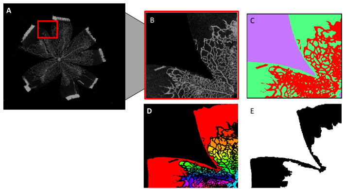
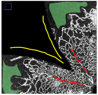
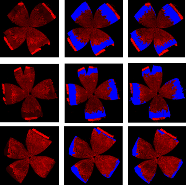
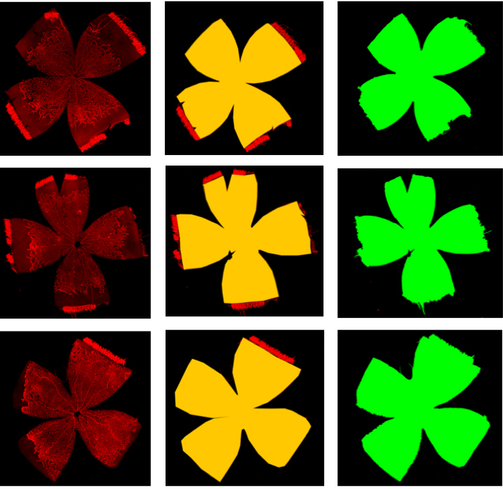
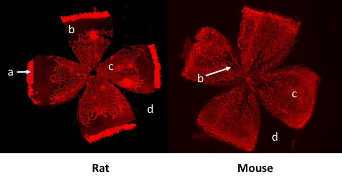
Within 6 h of birth, full-term pups born to Sprague Dawley rat dams that had undergone partial bilateral uterine artery ligation at embryonic day 19.5 were placed into a controlled oxygen environment (Oxycycler, BioSpherix, Parish, NY) at 50% oxygen for 48 h, followed by cycling between 10% and 50% oxygen every 24 h until day 15. The pups were then moved into room air until day 18.5. Ten lectin-stained retinal flat mounts were imaged in montage fashion at 10x magnification. Three masked human reviewers measured two parameters, total retinal area and peripheral avascular area, for each image using the ImageJ freehand selection tool. The outputs of each read were measured as number of pixels. The gold standard value for each image was the mean of the three human reads. Interrater agreement for the measurement of total retinal area, avascular area, and percent avascular area was calculated using type A intraclass correlation coefficients (ICCs) with a two-way random effects model. Automated avascular area identification (A3ID) is a method written in ImageJ Macro that is intended for use in the Fiji (Fiji is Just ImageJ) image processing platform. The input for A3ID is a rat retinal image, and the output is the avascular area (in pixels). A3ID utilizes a random forest classifier with a connected-components algorithm and post-processing filters for size and shape. A separate algorithm calculates the total retinal area. We compared the output of both algorithms to gold standard measurements by calculating ICCs, performing linear regression, and determining the Dice coefficients for both algorithms. We also constructed a Bland-Altman plot for A3ID output.
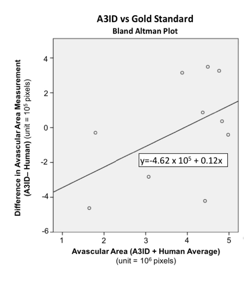
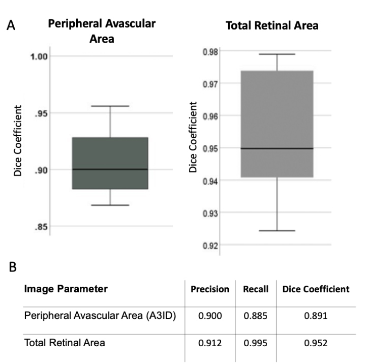
The ICC for percent peripheral avascular/total area between human readers was 0.995 (CI: 0.974-0.999), with p<0.001. The ICC between A3ID and the gold standard was calculated for three image parameters-avascular area: 0.974 (CI: 0.899-0.993), with p<0.001; total retinal area: 0.465 (CI: 0.0-0.851), with p=0.001; and the percent peripheral avascular/total area: 0.94 (CI: 0.326-0.989), with p<0.001. In the linear regression analysis, the slope for prediction of the gold standard percent peripheral avascular/total area from A3ID was 0.98, with R2=0.975. A3ID and the total retinal area algorithm achieve an average Dice coefficient of 0.891 and 0.952, respectively. The Bland-Altman analysis revealed a trend for computer underestimation of the peripheral avascular area in images with low peripheral avascular area and overestimation of peripheral avascular area in images with large peripheral avascular areas.
A3ID reliably predicts peripheral avascular area based on rat OIR retinal images. When the peripheral avascular area is particularly high or low, hand segmentation of images may be superior.
本研究的目的是创建一种算法,以实现大鼠氧诱导视网膜病变(OIR)模型图像中无血管区域分割过程的自动化、加速和标准化。
在出生后6小时内,将在胚胎第19.5天接受部分双侧子宫动脉结扎的Sprague Dawley大鼠母鼠所生的足月幼崽置于可控氧环境(Oxycycler,BioSpherix,Parish,纽约)中,在50%氧气浓度下放置48小时,然后每24小时在10%和50%氧气之间循环,直至第15天。然后将幼崽转移到室内空气中直至第18.5天。以10倍放大倍数对10个凝集素染色的视网膜平铺标本进行拼接成像。三名蒙面的人类审阅者使用ImageJ徒手选择工具为每个图像测量两个参数,即视网膜总面积和周边无血管区域。每次读数的输出以像素数衡量。每个图像的金标准值是三次人类读数的平均值。使用具有双向随机效应模型的A类组内相关系数(ICC)计算视网膜总面积、无血管区域和无血管区域百分比测量的审阅者间一致性。自动无血管区域识别(A3ID)是一种用ImageJ宏编写的方法,旨在用于Fiji(Fiji即Just ImageJ)图像处理平台。A3ID的输入是大鼠视网膜图像,输出是无血管区域(以像素为单位)。A3ID利用带有连通分量算法以及大小和形状后处理滤波器的随机森林分类器。一个单独的算法计算视网膜总面积。我们通过计算ICC、进行线性回归并确定两种算法的Dice系数,将两种算法的输出与金标准测量值进行比较。我们还为A3ID输出构建了Bland-Altman图。
人类审阅者之间周边无血管/总面积百分比的ICC为0.995(CI:0.974 - 0.999),p < 0.001。计算了A3ID与金标准之间针对三个图像参数的ICC——无血管区域:0.974(CI:0.899 - 0.993),p < 0.001;视网膜总面积:0.465(CI:0.0 - 0.851),p = 0.001;以及周边无血管/总面积百分比:0.94(CI:0.326 - 0.989),p < 0.001。在线性回归分析中,根据A3ID预测金标准周边无血管/总面积百分比的斜率为0.98,R² = 0.975。A3ID和视网膜总面积算法的平均Dice系数分别为0.891和0.952。Bland-Altman分析显示,在周边无血管区域低的图像中计算机有低估周边无血管区域的趋势,而在周边无血管区域大的图像中有高估周边无血管区域的趋势。
A3ID基于大鼠OIR视网膜图像可靠地预测周边无血管区域。当周边无血管区域特别高或低时,图像的手动分割可能更优。