• 文献检索
  • 文档翻译
  • 深度研究
  • 学术资讯
  • Suppr Zotero 插件Zotero 插件
  • 邀请有礼
  • 套餐&价格
  • 历史记录
应用&插件
Suppr Zotero 插件Zotero 插件浏览器插件Mac 客户端Windows 客户端微信小程序
定价
高级版会员购买积分包购买API积分包
服务
文献检索文档翻译深度研究API 文档MCP 服务
关于我们
关于 Suppr公司介绍联系我们用户协议隐私条款
关注我们

Suppr 超能文献

核心技术专利:CN118964589B侵权必究
粤ICP备2023148730 号-1Suppr @ 2026

文献检索

告别复杂PubMed语法,用中文像聊天一样搜索,搜遍4000万医学文献。AI智能推荐,让科研检索更轻松。

立即免费搜索

文件翻译

保留排版,准确专业,支持PDF/Word/PPT等文件格式,支持 12+语言互译。

免费翻译文档

深度研究

AI帮你快速写综述,25分钟生成高质量综述,智能提取关键信息,辅助科研写作。

立即免费体验

基于超高场 MP2RAGE 的多发性硬化病变的自动检测和分割。

Automated Detection and Segmentation of Multiple Sclerosis Lesions Using Ultra-High-Field MP2RAGE.

机构信息

Department of Radiology, Centre Hospitalier Universitaire Vaudois.

Signal Processing Laboratory (LTS 5), Ecole Polytechnique Fédérale de Lausanne, Lausanne.

出版信息

Invest Radiol. 2019 Jun;54(6):356-364. doi: 10.1097/RLI.0000000000000551.

DOI:10.1097/RLI.0000000000000551
PMID:30829941
原文链接:https://pmc.ncbi.nlm.nih.gov/articles/PMC6499666/
Abstract

OBJECTIVES

The aim of this study was to develop a new automated segmentation method of white matter (WM) and cortical multiple sclerosis (MS) lesions visible on magnetization-prepared 2 inversion-contrast rapid gradient echo (MP2RAGE) images acquired at 7 T MRI.

MATERIALS AND METHODS

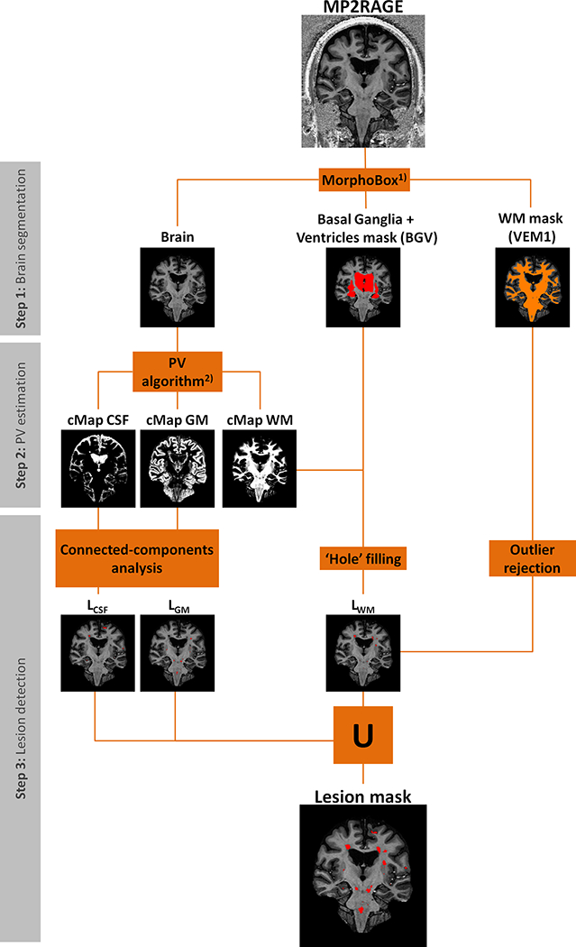
The proposed prototype (MSLAST [Multiple Sclerosis Lesion Analysis at Seven Tesla]) takes as input a single image contrast derived from the 7T MP2RAGE prototype sequence and is based on partial volume estimation and topological constraints. First, MSLAST performs a skull-strip of MP2RAGE images and computes tissue concentration maps for WM, gray matter (GM), and cerebrospinal fluid (CSF) using a partial volume model of tissues within each voxel. Second, MSLAST performs (1) connected-component analysis to GM and CSF concentration maps to classify small isolated components as MS lesions; (2) hole-filling in the WM concentration map to classify areas with low WM concentration surrounded by WM (ie, MS lesions); and (3) outlier rejection to the WM mask to improve the classification of small WM lesions. Third, MSLAST unifies the 3 maps obtained from 1, 2, and 3 processing steps to generate a global lesion mask.

RESULTS

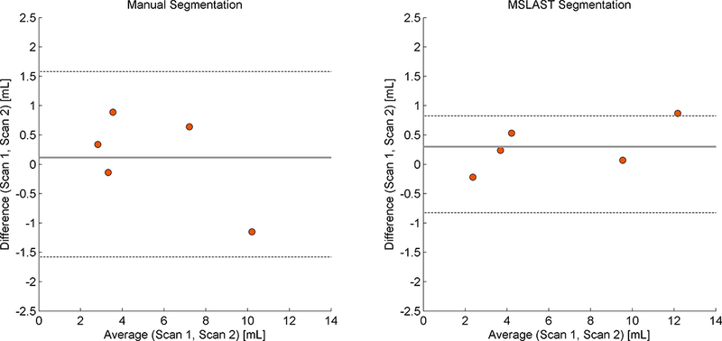
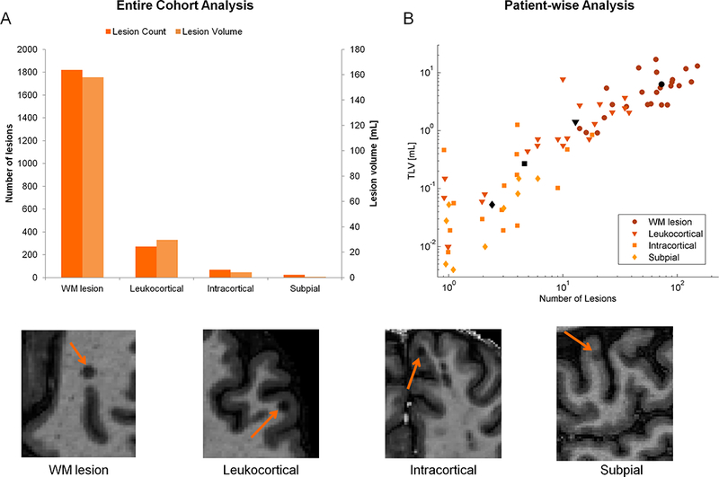
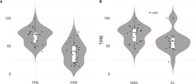
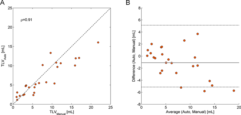
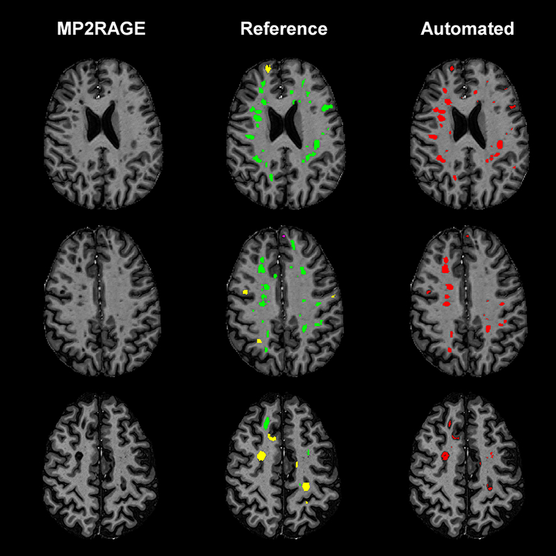
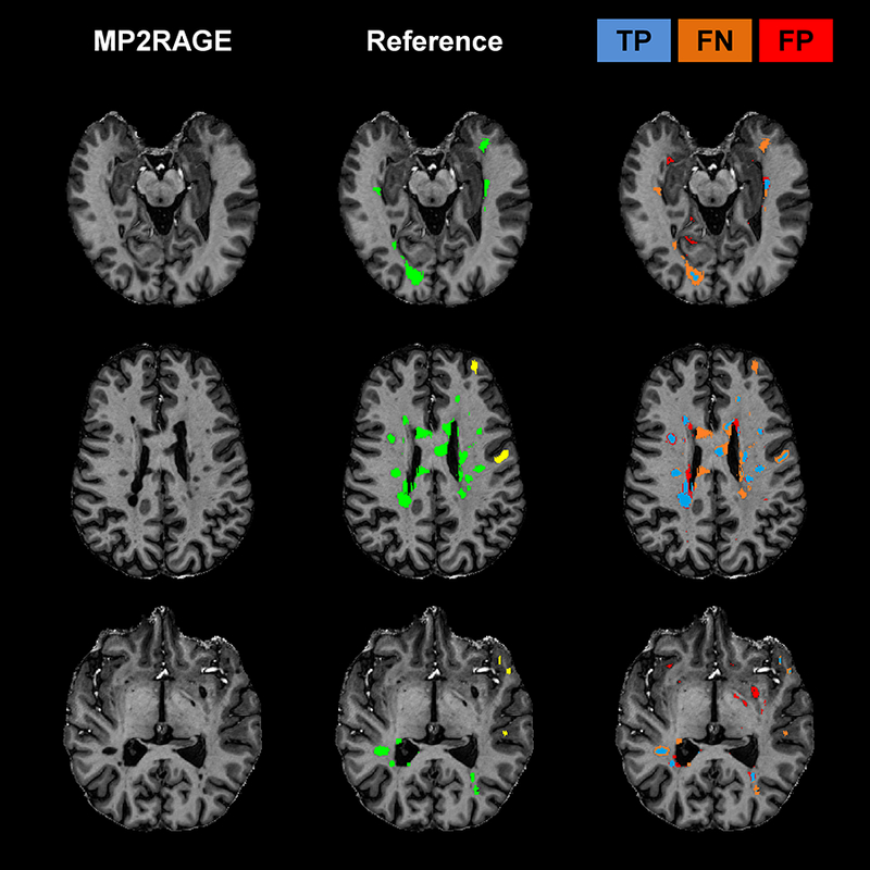
Quantitative and qualitative assessments were performed using MSLAST in 25 MS patients from 2 research centers. Overall, MSLAST detected a median of 71% of MS lesions, specifically 74% of WM and 58% of cortical lesions, when a minimum lesion size of 6 μL was considered. The median false-positive rate was 40%. When a 15 μL minimal lesions size was applied, which is the approximation of the minimal size recommended for 1.5/3 T images, the median detection rate was 80% for WM and 63% for cortical lesions, respectively, and the median false-positive rate was 33%. We observed high correlation between MSLAST and manual segmentations (Spearman rank correlation coefficient, ρ = 0.91), although MSLAST underestimated the total lesion volume (average difference of 1.1 mL), especially in patients with high lesion loads. MSLAST also showed good scan-rescan repeatability within the same session with an average absolute volume difference and F1 score of 0.38 ± 0.32 mL and 84%, respectively.

CONCLUSIONS

We propose a new methodology to facilitate the segmentation of WM and cortical MS lesions at 7 T MRI, our approach uses a single MP2RAGE scan and may be of special interest to clinicians and researchers.

摘要

目的

本研究旨在开发一种新的自动化分割方法,用于对 7T MRI 上采集的磁化准备 2 反转对比快速梯度回波(MP2RAGE)图像中可见的白质(WM)和皮质多发性硬化(MS)病变进行分割。

材料与方法

所提出的原型(MSLAST [七特斯拉多发性硬化病变分析])以单个图像对比度作为输入,该对比度来自于 7T MP2RAGE 原型序列,并基于部分体积估计和拓扑约束。首先,MSLAST 对 MP2RAGE 图像进行头骨剥离,并使用每个体素内组织的部分体积模型计算 WM、灰质(GM)和脑脊液(CSF)的组织浓度图。其次,MSLAST 执行(1)连通分量分析,以 GM 和 CSF 浓度图为分类依据,将小的孤立成分归类为 MS 病变;(2)在 WM 浓度图中进行空洞填充,以将 WM 周围低 WM 浓度的区域归类为 MS 病变;(3)对 WM 掩模进行异常值拒绝处理,以改善小 WM 病变的分类。第三,MSLAST 将 1、2 和 3 个处理步骤获得的 3 个图谱进行统一,生成全局病变掩模。

结果

在来自 2 个研究中心的 25 名 MS 患者中,使用 MSLAST 进行了定量和定性评估。总体而言,当考虑最小病变大小为 6μL 时,MSLAST 检测到的 MS 病变中位数为 71%,具体为 WM 病变 74%,皮质病变 58%。假阳性率中位数为 40%。当应用最小病变大小为 15μL 时(这是对 1.5/3T 图像推荐的最小病变大小的近似值),WM 病变的中位检出率分别为 80%,皮质病变的中位检出率为 63%,假阳性率中位数为 33%。我们观察到 MSLAST 与手动分割之间具有高度相关性(Spearman 秩相关系数,ρ=0.91),尽管 MSLAST 低估了总病变体积(平均差异 1.1mL),尤其是在病变负荷较高的患者中。MSLAST 还在同一会话内显示出良好的扫描-重扫可重复性,平均绝对体积差异和 F1 分数分别为 0.38±0.32mL 和 84%。

结论

我们提出了一种新的方法,用于促进 7T MRI 上 WM 和皮质 MS 病变的分割,我们的方法仅使用单个 MP2RAGE 扫描,这可能对临床医生和研究人员特别有兴趣。

https://cdn.ncbi.nlm.nih.gov/pmc/blobs/f27c/6499666/beec8840fb5b/nihms-1518257-f0007.jpg
https://cdn.ncbi.nlm.nih.gov/pmc/blobs/f27c/6499666/bd73de4dadd4/nihms-1518257-f0001.jpg
https://cdn.ncbi.nlm.nih.gov/pmc/blobs/f27c/6499666/e7d7d2b7e026/nihms-1518257-f0002.jpg
https://cdn.ncbi.nlm.nih.gov/pmc/blobs/f27c/6499666/5910ca4ffc63/nihms-1518257-f0003.jpg
https://cdn.ncbi.nlm.nih.gov/pmc/blobs/f27c/6499666/ce79c992674f/nihms-1518257-f0004.jpg
https://cdn.ncbi.nlm.nih.gov/pmc/blobs/f27c/6499666/8d478f37070b/nihms-1518257-f0005.jpg
https://cdn.ncbi.nlm.nih.gov/pmc/blobs/f27c/6499666/63e24a92ebfd/nihms-1518257-f0006.jpg
https://cdn.ncbi.nlm.nih.gov/pmc/blobs/f27c/6499666/beec8840fb5b/nihms-1518257-f0007.jpg

相似文献

1
Automated Detection and Segmentation of Multiple Sclerosis Lesions Using Ultra-High-Field MP2RAGE.基于超高场 MP2RAGE 的多发性硬化病变的自动检测和分割。
Invest Radiol. 2019 Jun;54(6):356-364. doi: 10.1097/RLI.0000000000000551.
2
Fluid and White Matter Suppression: New Sensitive 3 T Magnetic Resonance Imaging Contrasts for Cortical Lesion Detection in Multiple Sclerosis.液体和白质抑制:新的敏感 3T 磁共振成像对比在多发性硬化症中的皮质病变检测。
Invest Radiol. 2022 Sep 1;57(9):592-600. doi: 10.1097/RLI.0000000000000877. Epub 2022 May 3.
3
Multiple sclerosis cortical lesion detection with deep learning at ultra-high-field MRI.深度学习在超高场 MRI 下对多发性硬化皮质病变的检测。
NMR Biomed. 2022 Aug;35(8):e4730. doi: 10.1002/nbm.4730. Epub 2022 Mar 31.
4
Automated detection of white matter and cortical lesions in early stages of multiple sclerosis.多发性硬化早期白质和皮质病变的自动检测
J Magn Reson Imaging. 2016 Jun;43(6):1445-54. doi: 10.1002/jmri.25095. Epub 2015 Nov 25.
5
Advantages of fluid and white matter suppression (FLAWS) with MP2RAGE compared with double inversion recovery turbo spin echo (DIR-TSE) at 7T.7T 下 MP2RAGE 与双反转恢复涡轮自旋回波(DIR-TSE)相比的优势在于具有流体液和白质抑制(FLAWS)。
Eur J Radiol. 2019 Jul;116:160-164. doi: 10.1016/j.ejrad.2019.04.019. Epub 2019 Apr 29.
6
7T MPFLAIR versus MP2RAGE for Quantifying Lesion Volume in Multiple Sclerosis.7T MPFLAIR 与 MP2RAGE 定量多发性硬化病变体积的比较。
J Neuroimaging. 2020 Jul;30(4):531-536. doi: 10.1111/jon.12718. Epub 2020 Jun 22.
7
An Ultra-High Field Study of Cerebellar Pathology in Early Relapsing-Remitting Multiple Sclerosis Using MP2RAGE.一项使用MP2RAGE对早期复发缓解型多发性硬化症小脑病理进行的超高场研究。
Invest Radiol. 2017 May;52(5):265-273. doi: 10.1097/RLI.0000000000000338.
8
White matter lesion filling improves the accuracy of cortical thickness measurements in multiple sclerosis patients: a longitudinal study.白质病变填充可提高多发性硬化症患者皮质厚度测量的准确性:一项纵向研究。
BMC Neurosci. 2014 Sep 8;15:106. doi: 10.1186/1471-2202-15-106.
9
Improved Visualization of Cortical Lesions in Multiple Sclerosis Using 7T MP2RAGE.使用 7T MP2RAGE 提高多发性硬化症皮质病变的可视化效果。
AJNR Am J Neuroradiol. 2018 Mar;39(3):459-466. doi: 10.3174/ajnr.A5534. Epub 2018 Feb 8.
10
Inversion Recovery Susceptibility Weighted Imaging With Enhanced T2 Weighting at 3 T Improves Visualization of Subpial Cortical Multiple Sclerosis Lesions.3T 增强 T2 加权反转恢复敏感加权成像可提高脑皮层下多发性硬化病变的可视化效果。
Invest Radiol. 2020 Nov;55(11):727-735. doi: 10.1097/RLI.0000000000000698.

引用本文的文献

1
The use of 7T MRI in multiple sclerosis: review and consensus statement from the North American Imaging in Multiple Sclerosis Cooperative.7T磁共振成像在多发性硬化症中的应用:北美多发性硬化症协作组的综述与共识声明
Brain Commun. 2024 Oct 9;6(5):fcae359. doi: 10.1093/braincomms/fcae359. eCollection 2024.
2
Advanced MRI Techniques: Diagnosis and Follow-Up of Multiple Sclerosis.先进的磁共振成像技术:多发性硬化症的诊断与随访
Diagnostics (Basel). 2024 May 28;14(11):1120. doi: 10.3390/diagnostics14111120.
3
Present and future of the diagnostic work-up of multiple sclerosis: the imaging perspective.

本文引用的文献

1
Improved Visualization of Cortical Lesions in Multiple Sclerosis Using 7T MP2RAGE.使用 7T MP2RAGE 提高多发性硬化症皮质病变的可视化效果。
AJNR Am J Neuroradiol. 2018 Mar;39(3):459-466. doi: 10.3174/ajnr.A5534. Epub 2018 Feb 8.
2
Diagnosis of multiple sclerosis: 2017 revisions of the McDonald criteria.多发性硬化症的诊断:2017 年麦当劳标准修订版。
Lancet Neurol. 2018 Feb;17(2):162-173. doi: 10.1016/S1474-4422(17)30470-2. Epub 2017 Dec 21.
3
Improving automated multiple sclerosis lesion segmentation with a cascaded 3D convolutional neural network approach.
多发性硬化症的诊断工作现状和未来:影像学视角。
J Neurol. 2023 Mar;270(3):1286-1299. doi: 10.1007/s00415-022-11488-y. Epub 2022 Nov 24.
4
Predictive value of number and volume of demyelinating plaques in treatment response in patients with multiple sclerosis treated with INF-B.干扰素-β治疗的多发性硬化症患者中,脱髓鞘斑块数量和体积对治疗反应的预测价值。
Am J Neurodegener Dis. 2022 Apr 15;11(1):10-16. eCollection 2022.
5
Multiple sclerosis cortical lesion detection with deep learning at ultra-high-field MRI.深度学习在超高场 MRI 下对多发性硬化皮质病变的检测。
NMR Biomed. 2022 Aug;35(8):e4730. doi: 10.1002/nbm.4730. Epub 2022 Mar 31.
6
Automatic segmentation of white matter hyperintensities: validation and comparison with state-of-the-art methods on both Multiple Sclerosis and elderly subjects.自动分割脑白质高信号:在多发性硬化症和老年患者中验证和比较最先进的方法。
Neuroimage Clin. 2022;33:102940. doi: 10.1016/j.nicl.2022.102940. Epub 2022 Jan 10.
7
Imaging cortical multiple sclerosis lesions with ultra-high field MRI.超高场 MRI 成像皮质多发性硬化病变。
Neuroimage Clin. 2021;32:102847. doi: 10.1016/j.nicl.2021.102847. Epub 2021 Oct 6.
8
MRI with ultrahigh field strength and high-performance gradients: challenges and opportunities for clinical neuroimaging at 7 T and beyond.具有超高场强和高性能梯度的磁共振成像:7T及更高场强下临床神经成像面临的挑战与机遇
Eur Radiol Exp. 2021 Aug 26;5(1):35. doi: 10.1186/s41747-021-00216-2.
9
7T MRI Differentiates Remyelinated from Demyelinated Multiple Sclerosis Lesions.7T MRI 可区分脱髓鞘和复发性多发性硬化病变。
Ann Neurol. 2021 Oct;90(4):612-626. doi: 10.1002/ana.26194. Epub 2021 Sep 2.
10
An anomaly detection approach to identify chronic brain infarcts on MRI.一种用于在 MRI 上识别慢性脑梗死的异常检测方法。
Sci Rep. 2021 Apr 8;11(1):7714. doi: 10.1038/s41598-021-87013-4.
采用级联3D卷积神经网络方法改进自动多发性硬化病变分割
Neuroimage. 2017 Jul 15;155:159-168. doi: 10.1016/j.neuroimage.2017.04.034. Epub 2017 Apr 19.
4
Longitudinal multiple sclerosis lesion segmentation: Resource and challenge.纵向多发性硬化病变分割:资源与挑战。
Neuroimage. 2017 Mar 1;148:77-102. doi: 10.1016/j.neuroimage.2016.12.064. Epub 2017 Jan 11.
5
An Ultra-High Field Study of Cerebellar Pathology in Early Relapsing-Remitting Multiple Sclerosis Using MP2RAGE.一项使用MP2RAGE对早期复发缓解型多发性硬化症小脑病理进行的超高场研究。
Invest Radiol. 2017 May;52(5):265-273. doi: 10.1097/RLI.0000000000000338.
6
Neuroinflammatory component of gray matter pathology in multiple sclerosis.多发性硬化症中灰质病理的神经炎症成分
Ann Neurol. 2016 Nov;80(5):776-790. doi: 10.1002/ana.24791. Epub 2016 Oct 25.
7
Increased cortical grey matter lesion detection in multiple sclerosis with 7 T MRI: a post-mortem verification study.7T MRI 检测多发性硬化症皮质灰质病变的增加:一项死后验证研究。
Brain. 2016 May;139(Pt 5):1472-81. doi: 10.1093/brain/aww037. Epub 2016 Mar 8.
8
Deep 3D Convolutional Encoder Networks With Shortcuts for Multiscale Feature Integration Applied to Multiple Sclerosis Lesion Segmentation.具有捷径的深层 3D 卷积编码网络,用于多尺度特征集成,应用于多发性硬化病变分割。
IEEE Trans Med Imaging. 2016 May;35(5):1229-1239. doi: 10.1109/TMI.2016.2528821. Epub 2016 Feb 11.
9
Comparison of Routine Brain Imaging at 3 T and 7 T.3T和7T常规脑成像的比较
Invest Radiol. 2016 Aug;51(8):469-82. doi: 10.1097/RLI.0000000000000256.
10
Automated detection of white matter and cortical lesions in early stages of multiple sclerosis.多发性硬化早期白质和皮质病变的自动检测
J Magn Reson Imaging. 2016 Jun;43(6):1445-54. doi: 10.1002/jmri.25095. Epub 2015 Nov 25.