University Heart Center, Department of Cardiology, University Hospital Zurich, Raemistrasse 100, Zurich, Switzerland.
Division Neuropsychology, Department of Psychology, University of Zurich, Binzmuehlestrasse 14, Zurich, Switzerland.
Eur Heart J. 2019 Apr 14;40(15):1183-1187. doi: 10.1093/eurheartj/ehz068.
Takotsubo syndrome (TTS) is characterized by acute left ventricular dysfunction often triggered by emotional or physical stress. Severe activation of the sympathetic nervous system with catecholamine release caused by a dysfunctional limbic system has been proposed as a potential mechanism. We hypothesize that brain regions responsible for autonomic integration and/or limbic processing might be involved in the development of TTS. Here, we investigated alterations in resting state functional connectivity in TTS patients compared with healthy controls.
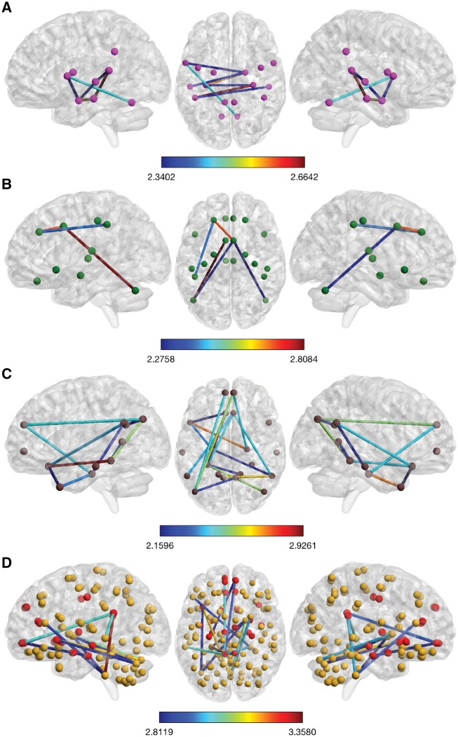
Using brain functional magnetic resonance imaging (fMRI), resting state functional connectivity has been assessed in 15 subjects with TTS and 39 healthy controls. Network-based statistical analyses were conducted to identify subnetworks with altered resting state functional connectivity. Sympathetic and parasympathetic networks have been constructed in addition to the default mode network and whole-brain network. We found parasympathetic- and sympathetic-associated subnetworks both showing reduced resting state functional connectivity in TTS patients compared with controls. Important brain regions constituting parasympathetic- and sympathetic-associated subnetworks included the amygdala, hippocampus, and insula as well as cingulate, parietal, temporal, and cerebellar regions. Additionally, the default mode network as well as limbic regions in the whole-brain analysis demonstrated reduced resting state functional connectivity in TTS, including the hippocampus, parahippocampal, and medial prefrontal regions.
For the first time, we demonstrate hypoconnectivity of central brain regions associated with autonomic functions and regulation of the limbic system in patients with TTS. These findings suggest that autonomic-limbic integration might play an important role in the pathophysiology and contribute to the understanding of TTS.
Takotsubo 综合征(TTS)的特征是左心室功能急性障碍,通常由情绪或身体应激引发。目前提出的潜在发病机制是由于边缘系统功能障碍导致交感神经系统的严重激活和儿茶酚胺释放。我们假设负责自主整合和/或边缘处理的脑区可能参与 TTS 的发生。在此,我们研究了 TTS 患者与健康对照者之间静息状态功能连接的变化。
使用脑功能磁共振成像(fMRI),我们评估了 15 例 TTS 患者和 39 名健康对照者的静息状态功能连接。进行了基于网络的统计分析,以识别静息状态功能连接改变的子网络。除了默认模式网络和全脑网络外,还构建了交感和副交感网络。我们发现,与对照组相比,TTS 患者的副交感和交感相关子网的静息状态功能连接均减弱。构成副交感和交感相关子网的重要脑区包括杏仁核、海马体和脑岛以及扣带回、顶叶、颞叶和小脑区域。此外,在全脑分析中,默认模式网络以及边缘区域的静息状态功能连接在 TTS 中减弱,包括海马体、旁海马体和内侧前额叶区域。
我们首次证明了 TTS 患者与自主功能和边缘系统调节相关的中枢脑区的连接减少。这些发现表明自主-边缘整合可能在病理生理学中起重要作用,并有助于理解 TTS。