• 文献检索
  • 文档翻译
  • 深度研究
  • 学术资讯
  • Suppr Zotero 插件Zotero 插件
  • 邀请有礼
  • 套餐&价格
  • 历史记录
应用&插件
Suppr Zotero 插件Zotero 插件浏览器插件Mac 客户端Windows 客户端微信小程序
定价
高级版会员购买积分包购买API积分包
服务
文献检索文档翻译深度研究API 文档MCP 服务
关于我们
关于 Suppr公司介绍联系我们用户协议隐私条款
关注我们

Suppr 超能文献

核心技术专利:CN118964589B侵权必究
粤ICP备2023148730 号-1Suppr @ 2026

文献检索

告别复杂PubMed语法,用中文像聊天一样搜索,搜遍4000万医学文献。AI智能推荐,让科研检索更轻松。

立即免费搜索

文件翻译

保留排版,准确专业,支持PDF/Word/PPT等文件格式,支持 12+语言互译。

免费翻译文档

深度研究

AI帮你快速写综述,25分钟生成高质量综述,智能提取关键信息,辅助科研写作。

立即免费体验

基质溶脂素-自分泌运动因子信号轴促进胰腺肿瘤进展。

A Stromal Lysolipid-Autotaxin Signaling Axis Promotes Pancreatic Tumor Progression.

机构信息

Cancer Research UK Beatson Institute, Glasgow, UK.

Institute of Cancer Sciences, University of Glasgow, Glasgow, UK.

出版信息

Cancer Discov. 2019 May;9(5):617-627. doi: 10.1158/2159-8290.CD-18-1212. Epub 2019 Mar 5.

DOI:10.1158/2159-8290.CD-18-1212
PMID:30837243
原文链接:https://pmc.ncbi.nlm.nih.gov/articles/PMC6497553/
Abstract

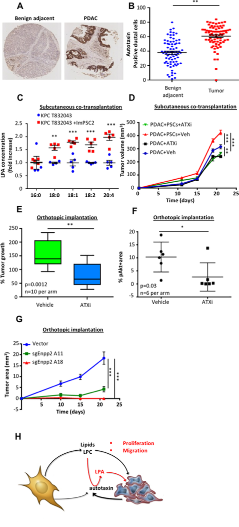
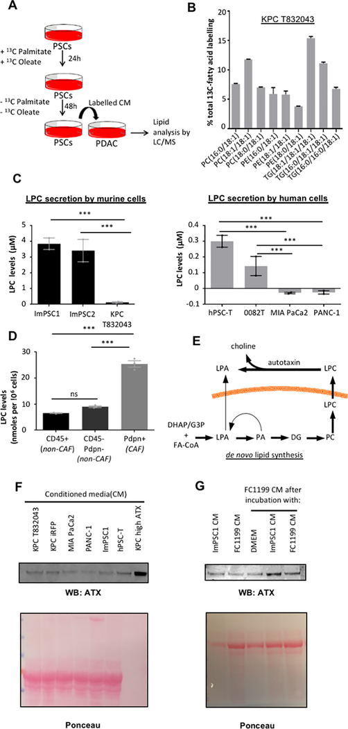
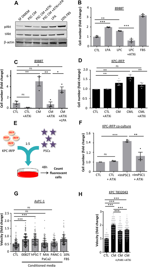
Pancreatic ductal adenocarcinoma (PDAC) develops a pronounced stromal response reflecting an aberrant wound-healing process. This stromal reaction features transdifferentiation of tissue-resident pancreatic stellate cells (PSC) into activated cancer-associated fibroblasts, a process induced by PDAC cells but of unclear significance for PDAC progression. Here, we show that PSCs undergo a dramatic lipid metabolic shift during differentiation in the context of pancreatic tumorigenesis, including remodeling of the intracellular lipidome and secretion of abundant lipids in the activated, fibroblastic state. Specifically, stroma-derived lysophosphatidylcholines support PDAC cell synthesis of phosphatidylcholines, key components of cell membranes, and also facilitate production of the potent wound-healing mediator lysophosphatidic acid (LPA) by the extracellular enzyme autotaxin, which is overexpressed in PDAC. The autotaxin-LPA axis promotes PDAC cell proliferation, migration, and AKT activation, and genetic or pharmacologic autotaxin inhibition suppresses PDAC growth . Our work demonstrates how PDAC cells exploit the local production of wound-healing mediators to stimulate their own growth and migration. SIGNIFICANCE: Our work highlights an unanticipated role for PSCs in producing the oncogenic LPA signaling lipid and demonstrates how PDAC tumor cells co-opt the release of wound-healing mediators by neighboring PSCs to promote their own proliferation and migration...

摘要

胰腺导管腺癌 (PDAC) 会产生明显的基质反应,反映出异常的伤口愈合过程。这种基质反应的特征是组织驻留的胰腺星状细胞 (PSC) 向激活的癌相关成纤维细胞转化,这一过程由 PDAC 细胞诱导,但对 PDAC 进展的意义尚不清楚。在这里,我们表明 PSC 在胰腺肿瘤发生过程中的分化过程中会经历剧烈的脂质代谢转变,包括细胞内脂质组的重塑和激活的成纤维状态下丰富脂质的分泌。具体而言,基质衍生的溶血磷脂酰胆碱支持 PDAC 细胞合成磷脂酰胆碱,这是细胞膜的关键成分,并且还促进细胞外酶自分泌酶产生强效伤口愈合介质溶血磷脂酸 (LPA),自分泌酶在 PDAC 中过表达。自分泌酶-LPA 轴促进 PDAC 细胞增殖、迁移和 AKT 激活,并且遗传或药理学自分泌酶抑制抑制 PDAC 生长。我们的工作证明了 PDAC 细胞如何利用局部产生的伤口愈合介质来刺激自身的生长和迁移。意义:我们的工作突出了 PSC 产生致癌 LPA 信号脂质的意外作用,并展示了 PDAC 肿瘤细胞如何利用邻近 PSC 释放的伤口愈合介质来促进自身的增殖和迁移。

https://cdn.ncbi.nlm.nih.gov/pmc/blobs/06e6/6497553/861e409377f2/nihms-1523395-f0004.jpg
https://cdn.ncbi.nlm.nih.gov/pmc/blobs/06e6/6497553/d503fc1ba743/nihms-1523395-f0001.jpg
https://cdn.ncbi.nlm.nih.gov/pmc/blobs/06e6/6497553/e95d1a266757/nihms-1523395-f0002.jpg
https://cdn.ncbi.nlm.nih.gov/pmc/blobs/06e6/6497553/bdfbd10a0187/nihms-1523395-f0003.jpg
https://cdn.ncbi.nlm.nih.gov/pmc/blobs/06e6/6497553/861e409377f2/nihms-1523395-f0004.jpg

相似文献

1
A Stromal Lysolipid-Autotaxin Signaling Axis Promotes Pancreatic Tumor Progression.基质溶脂素-自分泌运动因子信号轴促进胰腺肿瘤进展。
Cancer Discov. 2019 May;9(5):617-627. doi: 10.1158/2159-8290.CD-18-1212. Epub 2019 Mar 5.
2
Galectin-3 Mediates Tumor Cell-Stroma Interactions by Activating Pancreatic Stellate Cells to Produce Cytokines via Integrin Signaling.半乳糖凝集素-3 通过激活胰腺星状细胞通过整合素信号产生细胞因子来介导肿瘤细胞-基质相互作用。
Gastroenterology. 2018 Apr;154(5):1524-1537.e6. doi: 10.1053/j.gastro.2017.12.014. Epub 2017 Dec 21.
3
Pancreatic stellate cell secreted IL-6 stimulates STAT3 dependent invasiveness of pancreatic intraepithelial neoplasia and cancer cells.胰腺星状细胞分泌的白细胞介素-6刺激胰腺上皮内瘤变和癌细胞的STAT3依赖性侵袭。
Oncotarget. 2016 Oct 4;7(40):65982-65992. doi: 10.18632/oncotarget.11786.
4
Positive feedback in Cav-1-ROS signalling in PSCs mediates metabolic coupling between PSCs and tumour cells.肝星状细胞中 Cav-1-ROS 信号的正反馈调节介导了肝星状细胞与肿瘤细胞之间的代谢偶联。
J Cell Mol Med. 2020 Aug;24(16):9397-9408. doi: 10.1111/jcmm.15596. Epub 2020 Jul 7.
5
The role of S100A9 in the interaction between pancreatic ductal adenocarcinoma cells and stromal cells.S100A9 在胰腺导管腺癌细胞与基质细胞相互作用中的作用。
Cancer Immunol Immunother. 2022 Mar;71(3):705-718. doi: 10.1007/s00262-021-03026-y. Epub 2021 Aug 10.
6
Paracrine IL-6 signaling mediates the effects of pancreatic stellate cells on epithelial-mesenchymal transition via Stat3/Nrf2 pathway in pancreatic cancer cells.旁分泌 IL-6 信号通过 Stat3/Nrf2 通路介导胰腺星状细胞对胰腺癌细胞上皮间质转化的影响。
Biochim Biophys Acta Gen Subj. 2017 Feb;1861(2):296-306. doi: 10.1016/j.bbagen.2016.10.006. Epub 2016 Oct 14.
7
Periostin promotes the chemotherapy resistance to gemcitabine in pancreatic cancer.骨膜蛋白促进胰腺癌对吉西他滨的化疗耐药性。
Tumour Biol. 2016 Nov;37(11):15283-15291. doi: 10.1007/s13277-016-5321-6. Epub 2016 Sep 30.
8
Activated pancreatic stellate cells sequester CD8+ T cells to reduce their infiltration of the juxtatumoral compartment of pancreatic ductal adenocarcinoma.活化的胰腺星状细胞将 CD8+T 细胞隔离,以减少其浸润胰腺导管腺癌的瘤周区域。
Gastroenterology. 2013 Nov;145(5):1121-32. doi: 10.1053/j.gastro.2013.07.025. Epub 2013 Jul 25.
9
Reprogramming pancreatic stellate cells via p53 activation: A putative target for pancreatic cancer therapy.通过激活 p53 重编程胰腺星状细胞:胰腺癌治疗的一个潜在靶点。
PLoS One. 2017 Dec 6;12(12):e0189051. doi: 10.1371/journal.pone.0189051. eCollection 2017.
10
Inhibiting tumor necrosis factor-alpha diminishes desmoplasia and inflammation to overcome chemoresistance in pancreatic ductal adenocarcinoma.抑制肿瘤坏死因子-α可减轻促结缔组织增生反应和炎症,从而克服胰腺导管腺癌的化疗耐药性。
Oncotarget. 2016 Dec 6;7(49):81110-81122. doi: 10.18632/oncotarget.13212.

引用本文的文献

1
Role of metabolic reprogramming of cancer‑associated fibroblasts in tumor development and progression (Review).癌症相关成纤维细胞的代谢重编程在肿瘤发生和进展中的作用(综述)
Int J Oncol. 2025 Nov;67(5). doi: 10.3892/ijo.2025.5796. Epub 2025 Aug 29.
2
Cannabidiol Is Associated with Improved Survival in Pancreatic Cancer and Modulation of Bile Acids and Gut Microbiota.大麻二酚与胰腺癌患者生存率提高以及胆汁酸和肠道微生物群的调节有关。
Int J Mol Sci. 2025 Aug 10;26(16):7733. doi: 10.3390/ijms26167733.
3
Domestication and feedback: bidirectional hijacking in pancreatic ductal adenocarcinoma microenvironment.

本文引用的文献

1
Stellate Cells in Tissue Repair, Inflammation, and Cancer.星状细胞在组织修复、炎症和癌症中的作用。
Annu Rev Cell Dev Biol. 2018 Oct 6;34:333-355. doi: 10.1146/annurev-cellbio-100617-062855. Epub 2018 Jul 20.
2
Adipocyte-Derived Lipids Mediate Melanoma Progression via FATP Proteins.脂肪细胞衍生的脂质通过 FATP 蛋白介导黑色素瘤的进展。
Cancer Discov. 2018 Aug;8(8):1006-1025. doi: 10.1158/2159-8290.CD-17-1371. Epub 2018 Jun 14.
3
Reprogramming pancreatic stellate cells via p53 activation: A putative target for pancreatic cancer therapy.
驯化与反馈:胰腺导管腺癌微环境中的双向劫持
Front Immunol. 2025 Aug 11;16:1585858. doi: 10.3389/fimmu.2025.1585858. eCollection 2025.
4
Biophysical and biochemical signatures of pancreatic stellate cell activation: insights into mechano-metabolic signalling from atomic force microscopy and Raman spectroscopy.胰腺星状细胞激活的生物物理和生化特征:基于原子力显微镜和拉曼光谱对机械代谢信号传导的见解
Cell Commun Signal. 2025 Aug 4;23(1):363. doi: 10.1186/s12964-025-02354-1.
5
What's on the menu?: metabolic constraints in the pancreatic tumor microenvironment.菜单上有什么?:胰腺肿瘤微环境中的代谢限制
J Clin Invest. 2025 Jul 15;135(14). doi: 10.1172/JCI191940.
6
Integrated spatial omics of metabolic reprogramming and the tumor microenvironment in pancreatic cancer.胰腺癌中代谢重编程与肿瘤微环境的综合空间组学
iScience. 2025 May 15;28(6):112681. doi: 10.1016/j.isci.2025.112681. eCollection 2025 Jun 20.
7
Tumor-stromal metabolic crosstalk in pancreatic cancer.胰腺癌中的肿瘤-基质代谢串扰
Trends Cell Biol. 2025 May 26. doi: 10.1016/j.tcb.2025.04.007.
8
Benign non-immune cells in tumor microenvironment.肿瘤微环境中的良性非免疫细胞。
Front Immunol. 2025 Apr 3;16:1561577. doi: 10.3389/fimmu.2025.1561577. eCollection 2025.
9
Microenvironmental arginine restriction sensitizes pancreatic cancers to polyunsaturated fatty acids by suppression of lipid synthesis.微环境精氨酸限制通过抑制脂质合成使胰腺癌对多不饱和脂肪酸敏感。
bioRxiv. 2025 Mar 13:2025.03.10.642426. doi: 10.1101/2025.03.10.642426.
10
Cancer-Associated Fibroblasts Foster a High-Lactate Microenvironment to Drive Perineural Invasion in Pancreatic Cancer.癌症相关成纤维细胞营造高乳酸微环境以驱动胰腺癌的神经周围浸润。
Cancer Res. 2025 Jun 16;85(12):2199-2217. doi: 10.1158/0008-5472.CAN-24-3173.
通过激活 p53 重编程胰腺星状细胞:胰腺癌治疗的一个潜在靶点。
PLoS One. 2017 Dec 6;12(12):e0189051. doi: 10.1371/journal.pone.0189051. eCollection 2017.
4
Distinct populations of inflammatory fibroblasts and myofibroblasts in pancreatic cancer.胰腺癌中不同的炎性成纤维细胞和肌成纤维细胞群体。
J Exp Med. 2017 Mar 6;214(3):579-596. doi: 10.1084/jem.20162024. Epub 2017 Feb 23.
5
Direct evidence for cancer-cell-autonomous extracellular protein catabolism in pancreatic tumors.胰腺肿瘤中癌细胞自主进行细胞外蛋白质分解代谢的直接证据。
Nat Med. 2017 Feb;23(2):235-241. doi: 10.1038/nm.4256. Epub 2016 Dec 26.
6
Pancreatic stellate cells support tumour metabolism through autophagic alanine secretion.胰腺星状细胞通过自噬性丙氨酸分泌来支持肿瘤代谢。
Nature. 2016 Aug 25;536(7617):479-83. doi: 10.1038/nature19084. Epub 2016 Aug 10.
7
Regulation and Functions of Autophagic Lipolysis.自噬性脂肪分解的调节与功能
Trends Endocrinol Metab. 2016 Oct;27(10):696-705. doi: 10.1016/j.tem.2016.06.003. Epub 2016 Jun 27.
8
Oncogenic KRAS Regulates Tumor Cell Signaling via Stromal Reciprocation.致癌性KRAS通过基质相互作用调节肿瘤细胞信号传导。
Cell. 2016 May 5;165(4):910-20. doi: 10.1016/j.cell.2016.03.029. Epub 2016 Apr 14.
9
Tumor microenvironment derived exosomes pleiotropically modulate cancer cell metabolism.肿瘤微环境衍生的外泌体多效性地调节癌细胞代谢。
Elife. 2016 Feb 27;5:e10250. doi: 10.7554/eLife.10250.
10
Stromal biology and therapy in pancreatic cancer: a changing paradigm.胰腺癌中的基质生物学和治疗:范式转变。
Gut. 2015 Sep;64(9):1476-84. doi: 10.1136/gutjnl-2015-309304. Epub 2015 May 20.