Cardiovascular Imaging Research Group, Heart and Vascular Center, Semmelweis University, 68. Varosmajor street, Budapest, Hungary.
Department of Internal Medicine and Cardiovascular Center, Seoul National University Hospital, 101 Daehang-ro, Chongno-gu, Seoul, Republic of Korea.
Eur Heart J Cardiovasc Imaging. 2019 Nov 1;20(11):1250-1258. doi: 10.1093/ehjci/jez033.
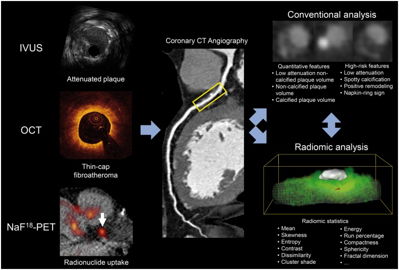
Identification of invasive and radionuclide imaging markers of coronary plaque vulnerability by a single, widely available non-invasive technique may provide the opportunity to identify vulnerable plaques and vulnerable patients in broad populations. Our aim was to assess whether radiomic analysis outperforms conventional assessment of coronary computed tomography angiography (CTA) images to identify invasive and radionuclide imaging markers of plaque vulnerability.
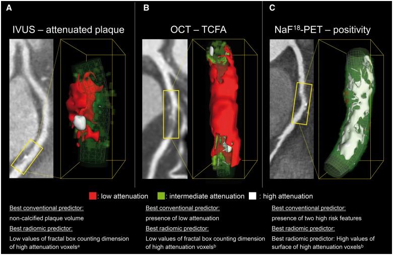
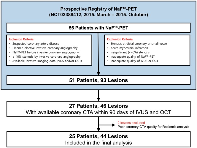
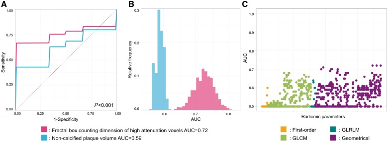
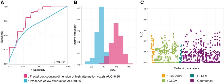
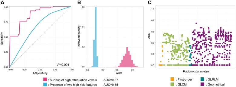
We prospectively included patients who underwent coronary CTA, sodium-fluoride positron emission tomography (NaF18-PET), intravascular ultrasound (IVUS), and optical coherence tomography (OCT). We assessed seven conventional plaque features and calculated 935 radiomic parameters from CTA images. In total, 44 plaques of 25 patients were analysed. The best radiomic parameters significantly outperformed the best conventional CT parameters to identify attenuated plaque by IVUS [fractal box counting dimension of high attenuation voxels vs. non-calcified plaque volume, area under the curve (AUC): 0.72, confidence interval (CI): 0.65-0.78 vs. 0.59, CI: 0.57-0.62; P < 0.001], thin-cap fibroatheroma by OCT (fractal box counting dimension of high attenuation voxels vs. presence of low attenuation voxels, AUC: 0.80, CI: 0.72-0.88 vs. 0.66, CI: 0.58-0.73; P < 0.001), and NaF18-positivity (surface of high attenuation voxels vs. presence of two high-risk features, AUC: 0.87, CI: 0.82-0.91 vs. 0.65, CI: 0.64-0.66; P < 0.001).
Coronary CTA radiomics identified invasive and radionuclide imaging markers of plaque vulnerability with good to excellent diagnostic accuracy, significantly outperforming conventional quantitative and qualitative high-risk plaque features. Coronary CTA radiomics may provide a more accurate tool to identify vulnerable plaques compared with conventional methods. Further larger population studies are warranted.
通过单一、广泛可用的无创技术识别冠状动脉斑块易损性的侵袭性和放射性核素成像标志物,可能有机会在广泛的人群中识别易损斑块和易损患者。我们的目的是评估放射组学分析是否优于冠状动脉计算机断层扫描血管造影(CTA)图像的常规评估,以识别斑块易损性的侵袭性和放射性核素成像标志物。
我们前瞻性纳入了接受冠状动脉 CTA、氟化钠正电子发射断层扫描(NaF18-PET)、血管内超声(IVUS)和光学相干断层扫描(OCT)检查的患者。我们评估了 7 种传统斑块特征,并从 CTA 图像中计算了 935 个放射组学参数。总共分析了 25 名患者的 44 个斑块。最佳放射组学参数显著优于最佳传统 CT 参数,可通过 IVUS 识别衰减斑块[高衰减体素分形盒计数维度与非钙化斑块体积,曲线下面积(AUC):0.72,置信区间(CI):0.65-0.78 与 0.59,CI:0.57-0.62;P<0.001]、OCT 中的薄帽纤维粥样斑块(高衰减体素分形盒计数维度与低衰减体素存在,AUC:0.80,CI:0.72-0.88 与 0.66,CI:0.58-0.73;P<0.001)和 NaF18 阳性(高衰减体素表面积与两个高危特征存在,AUC:0.87,CI:0.82-0.91 与 0.65,CI:0.64-0.66;P<0.001)。
冠状动脉 CTA 放射组学以良好到极好的诊断准确性识别了斑块易损性的侵袭性和放射性核素成像标志物,明显优于传统的定量和定性高危斑块特征。与传统方法相比,冠状动脉 CTA 放射组学可能提供一种更准确的识别易损斑块的工具。需要进一步进行更大规模的人群研究。