Masuda Naonori, Tsujinaka Hiroki, Hirai Hiromasa, Yamashita Mariko, Ueda Tetsuo, Ogata Nahoko
Department of Ophthalmology, Nara Medical University, 840 Shijo-cho, Kashihara, 634-8522, Japan.
BMC Ophthalmol. 2019 Mar 8;19(1):70. doi: 10.1186/s12886-019-1076-3.
Amyloid beta (Aβ) is a constituent of drusen that is a common sign of age-related macular degeneration (AMD). The purpose of this study was to investigate the effect of Aβ on human retinal pigment epithelial (RPE) cells in culture.
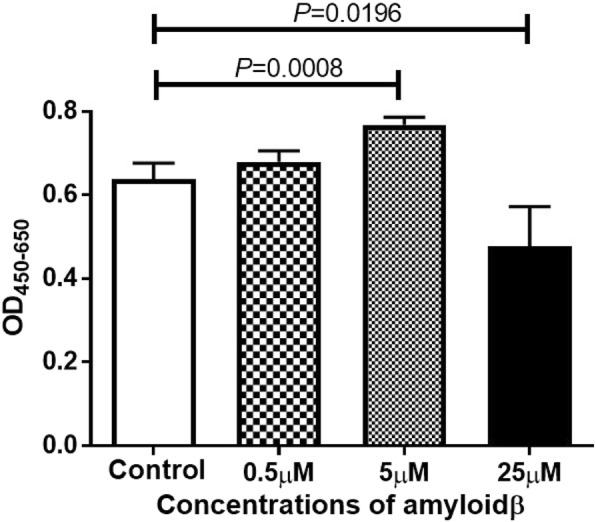
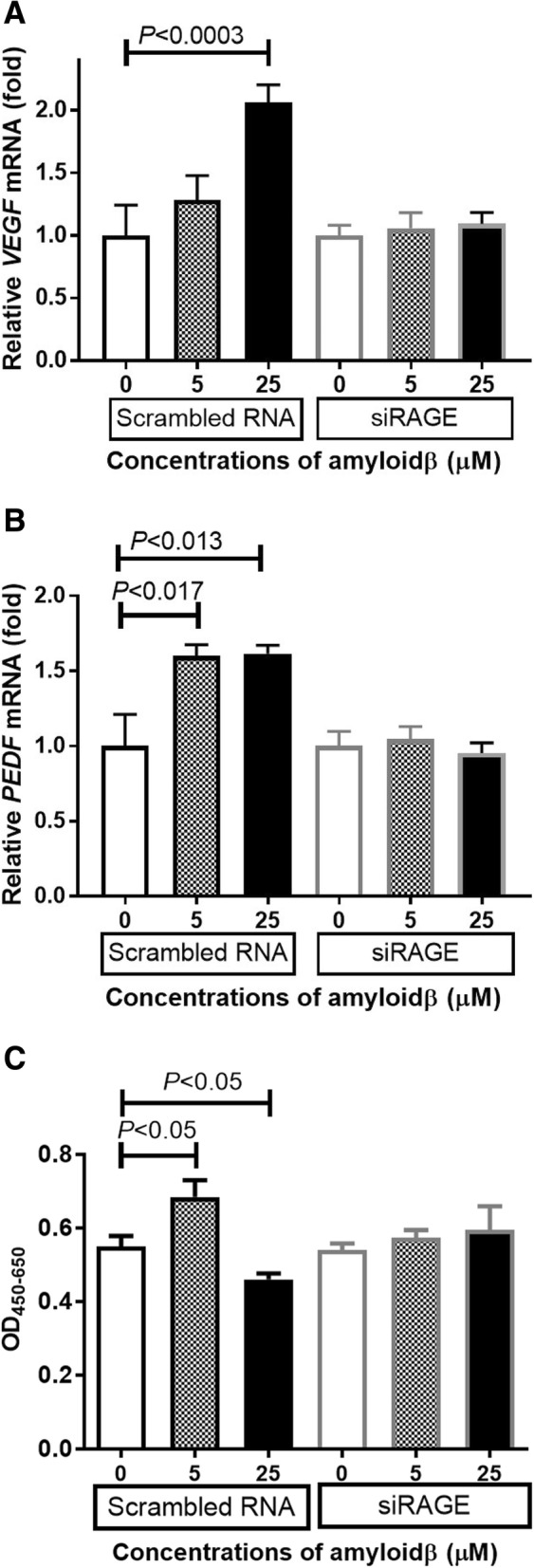
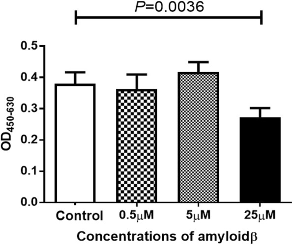
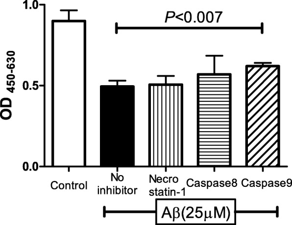
Cells from a human RPE cell line (ARPE-19) were exposed to 0 to 25 μM of Aβ 1-40 for 48 h, and the number of living cells was determined by WST-8 cleavage. Replicative DNA synthesis was measured by the incorporation of 5'-bromo-2'-deoxyuridine. The cell death pathway was investigated by the WST-8 cleavage assay after the addition of caspase-9 inhibitor, an anti-apoptotic factor. Real-time qRT-PCR was performed using Aβ-exposed cellular RNA to determine the level of vascular endothelial growth factor (VEGF)-A and pigment epithelium derived factor (PEDF). To determine the effect of receptor-for-advanced glycation end products (RAGE), the siRNA for RAGE was inserted into ARPE-19 treated with Aβ, and the levels of expression of VEGF-A and PEDF were determined.
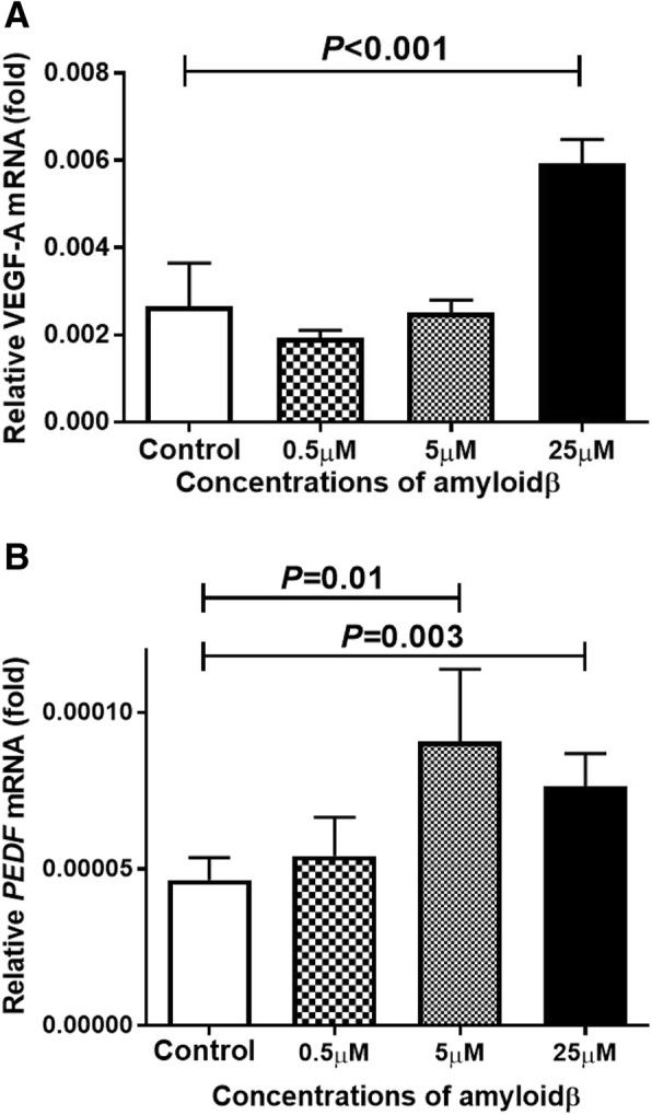
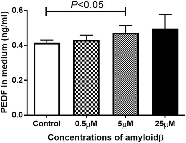
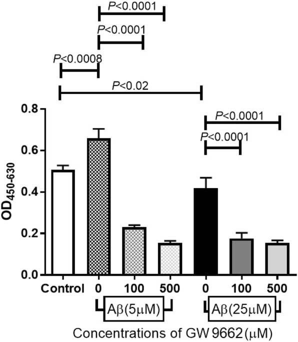
The number of living ARPE-19 cells was increased by exposure to 5 μM Aβ but was decreased by exposure to 25 μM of Aβ. Replicative DNA synthesis by ARPE-19 cells exposed to 25 μM of Aβ was significantly decreased indicating that 25 μM of Aβ inhibited cell proliferation. Real-time RT-PCR showed that the level of the mRNA of PEDF was increased by exposure to 5 μM Aβ, and the levels of the mRNAs of PEDF and VEGF-A were also increased by exposure to 25 μM Aβ. The addition of an inhibitor of caspase-9 blocked the decrease the number of ARPE-19 cells exposed to 25 μM Aβ. Exposure to si-RAGE attenuated the increase of VEGF-A and PEDF mRNA expression in ARPE-19 exposed to Aβ.
Exposure of ARPE-19 cells to low concentrations of Aβ increases the level of PEDF which then inhibits the apoptosis of ARPE-19 cells leading to RPE cell proliferation. Exposure to high concentrations of Aβ induces RPE cell death and enhances the expression of the mRNA of VEGF-A in RPE cells. The Aβ-RAGE pathway may lead to the expression VEGF-A and PEDF in RPE cells. These results suggest that Aβ is strongly related to the pathogenesis of choroidal neovascularization.
β淀粉样蛋白(Aβ)是玻璃膜疣的一种成分,玻璃膜疣是年龄相关性黄斑变性(AMD)的常见体征。本研究的目的是调查Aβ对培养的人视网膜色素上皮(RPE)细胞的影响。
将人RPE细胞系(ARPE-19)的细胞暴露于0至25μM的Aβ1-40中48小时,通过WST-8裂解测定活细胞数量。通过掺入5'-溴-2'-脱氧尿苷测量复制性DNA合成。在加入半胱天冬酶-9抑制剂(一种抗凋亡因子)后,通过WST-8裂解试验研究细胞死亡途径。使用暴露于Aβ的细胞RNA进行实时定量逆转录聚合酶链反应(qRT-PCR),以确定血管内皮生长因子(VEGF)-A和色素上皮衍生因子(PEDF)的水平。为了确定晚期糖基化终末产物受体(RAGE)的作用,将RAGE的小干扰RNA(siRNA)导入用Aβ处理的ARPE-19细胞中,并测定VEGF-A和PEDF的表达水平。
暴露于5μM Aβ可增加ARPE-19活细胞数量,但暴露于25μM Aβ则使其减少。暴露于25μM Aβ的ARPE-19细胞的复制性DNA合成显著减少,表明25μM Aβ抑制细胞增殖。实时逆转录聚合酶链反应(RT-PCR)显示,暴露于5μM Aβ可使PEDF的mRNA水平升高,暴露于25μM Aβ也可使PEDF和VEGF-A的mRNA水平升高。加入半胱天冬酶-9抑制剂可阻止暴露于25μM Aβ的ARPE-19细胞数量的减少。暴露于小干扰RNA-RAGE可减弱暴露于Aβ的ARPE-19细胞中VEGF-A和PEDF mRNA表达的增加。
ARPE-19细胞暴露于低浓度Aβ可增加PEDF水平,进而抑制ARPE-19细胞凋亡,导致RPE细胞增殖。暴露于高浓度Aβ可诱导RPE细胞死亡,并增强RPE细胞中VEGF-A mRNA的表达。Aβ-RAGE途径可能导致RPE细胞中VEGF-A和PEDF的表达。这些结果表明,Aβ与脉络膜新生血管形成的发病机制密切相关。