R&D Center, Lutronic Corporation, Goyang, Korea.
Global Center, Lutronic Corporation, Billerica, MA, USA.
Sci Rep. 2019 Mar 12;9(1):4186. doi: 10.1038/s41598-019-41021-7.
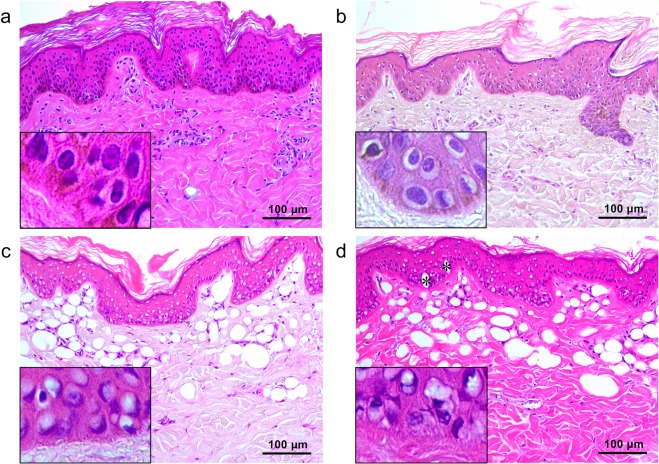
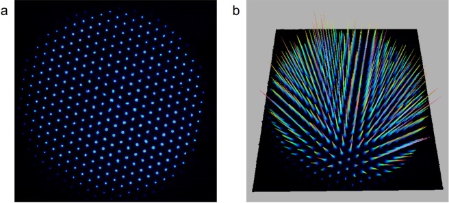
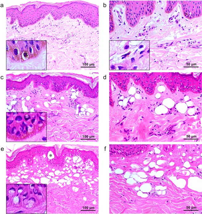
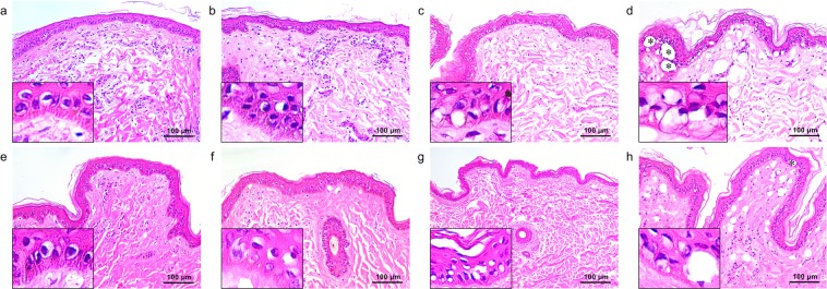
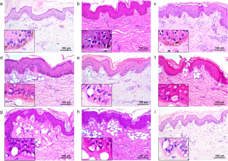
Optical pulses from picosecond lasers can be delivered to the skin as single, flat-top beams or fractionated beams using a beam splitter or microlens array (MLA). In this study, picosecond neodymium:yttrium aluminum garnet laser treatment using a single flat-top beam and an MLA-type beam at the wavelengths of 532 nm and 1,064 nm were delivered on ex vivo genotype-regulated, pigmented micropig skin. Skin specimens were obtained immediately after treatment and microscopically analyzed. Single flat-top beam treatment at a wavelength of 532 nm and a fluence of 0.05-J/cm reduced melanin pigments in epidermal keratinocytes and melanocytes, compared to untreated controls. Additionally, 0.1 J/cm- and 1.3 J/cm-fluenced laser treatment at 532 nm elicited noticeable vacuolation of keratinocytes and melanocytes within all epidermal layers. Single flat-top beam picosecond laser treatment at a wavelength of 1,064 nm and a fluence of 0.18 J/cm also reduced melanin pigments in keratinocytes and melanocytes. Treatment at 1,064-nm and fluences of 1.4 J/cm and 2.8 J/cm generated increasing degrees of vacuolated keratinocytes and melanocytes. Meanwhile, 532- and 1,064-nm MLA-type, picosecond laser treatment elicited fractionated zones of laser-induced micro-vacuolization in the epidermis and dermis. Therein, the sizes and degrees of tissue reactions differed according to wavelength, fluence, and distance between the microlens and skin.
皮秒激光的脉冲光可以通过分束器或微透镜阵列 (MLA) 传输到皮肤,形成单个平顶光束或分割光束。在这项研究中,使用波长为 532nm 和 1064nm 的单个平顶光束和 MLA 型光束对 ex vivo 基因型调控的色素性小母猪皮肤进行了皮秒掺钕钇铝石榴石激光处理。治疗后立即获得皮肤标本并进行显微镜分析。与未处理的对照组相比,532nm 波长和 0.05-J/cm 剂量的单一平顶光束处理降低了表皮角质形成细胞和黑素细胞中的黑色素色素。此外,532nm 波长的 0.1J/cm 和 1.3J/cm 激光处理引起了所有表皮层中角质形成细胞和黑素细胞的明显空泡化。1064nm 波长和 0.18J/cm 剂量的单一平顶光束皮秒激光处理也降低了角质形成细胞和黑素细胞中的黑色素色素。1064nm 波长和 1.4J/cm 和 2.8J/cm 的处理剂量产生了程度不同的空泡化角质形成细胞和黑素细胞。同时,532nm 和 1064nm 的 MLA 型皮秒激光处理在表皮和真皮中引发了分割的激光诱导微空化区。其中,根据波长、剂量和微透镜与皮肤之间的距离,组织反应的大小和程度有所不同。