Balu Mihaela, Lentsch Griffin, Korta Dorota Z, König Karsten, Kelly Kristen M, Tromberg Bruce J, Zachary Christopher B
Beckman Laser Institute, Laser Microbeam and Medical Program, University of California Irvine, Irvine, California, 92612.
Department of Dermatology, University of California, Irvine, California, 92697.
Lasers Surg Med. 2017 Aug;49(6):555-562. doi: 10.1002/lsm.22655. Epub 2017 Mar 23.
Improvements in skin appearance resulting from treatment with fractionated picosecond-lasers have been noted, but optimizing the treatment efficacy depends on a thorough understanding of the specific skin response. The development of non-invasive laser imaging techniques in conjunction with laser therapy can potentially provide feedback for guidance and optimizing clinical outcome.
The purpose of this study was to demonstrate the capability of multiphoton microscopy (MPM), a high-resolution, label-free imaging technique, to characterize in vivo the skin response to a fractionated non-ablative picosecond-laser treatment.
DESIGN, SETTING, AND PARTICIPANTS: Two areas on the arm of a volunteer were treated with a fractionated picosecond laser at the Dermatology Clinic, UC Irvine. The skin response to treatment was imaged in vivo with a clinical MPM-based tomograph at 3 hours and 24 hours after treatment and seven additional time points over a 4-week period.
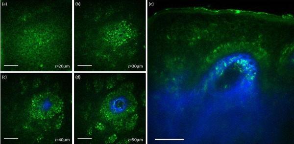
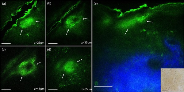
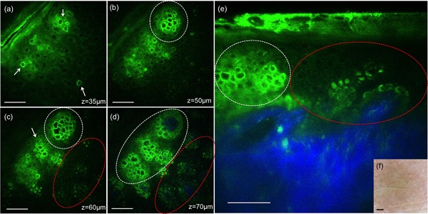
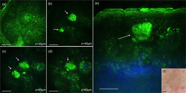
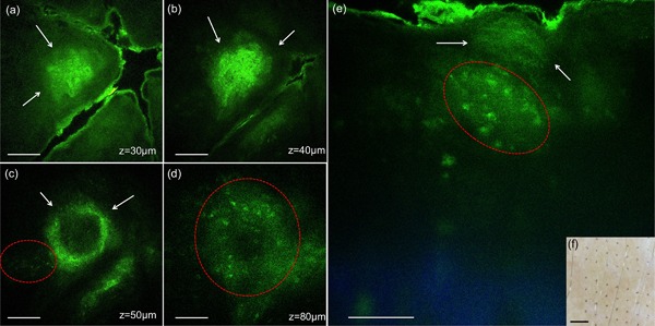
MPM revealed micro-injuries present in the epidermis. Pigmented cells were particularly damaged in the process, suggesting that melanin is likely the main absorber for laser induced optical breakdown.
Damaged individual cells were distinguished as early as 3 hours post pico-laser treatment with the 532 nm wavelength, and 24 hours post-treatment with both 532 and 1064 nm wavelengths. At later time points, clusters of cellular necrotic debris were imaged across the treated epidermis. After 24 hours of treatment, inflammatory cells were imaged in the proximity of epidermal micro-injuries. The epidermal injuries were exfoliated over a 4-week period.
This observational and descriptive pilot study demonstrates that in vivo MPM imaging can be used non-invasively to provide label-free contrast for describing changes in human skin following a fractionated non-ablative laser treatment. The results presented in this study represent the groundwork for future longitudinal investigations on an expanded number of subjects to understand the response to treatment in different skin types with different laser parameters, critical factors in optimizing treatment outcome. Lasers Surg. Med. 49:555-562, 2017. © 2017 Wiley Periodicals, Inc.
已注意到采用分馏皮秒激光治疗可改善皮肤外观,但要优化治疗效果,取决于对特定皮肤反应的透彻理解。结合激光疗法开发非侵入性激光成像技术可能为指导治疗和优化临床结果提供反馈。
本研究的目的是证明多光子显微镜(MPM)这一高分辨率、无标记成像技术在体内表征皮肤对分馏非剥脱性皮秒激光治疗反应的能力。
设计、地点和参与者:在加州大学欧文分校皮肤科诊所,用分馏皮秒激光对一名志愿者手臂上的两个区域进行治疗。在治疗后3小时和24小时以及4周内的另外七个时间点,使用基于临床MPM的断层扫描仪对皮肤的治疗反应进行体内成像。
MPM显示表皮存在微损伤。在此过程中色素细胞尤其受损,这表明黑色素可能是激光诱导光学击穿的主要吸收体。
在用532纳米波长的皮秒激光治疗后3小时以及用532和1064纳米波长治疗后24小时,就可最早区分出受损的单个细胞。在随后的时间点,在整个治疗过的表皮中可成像出细胞坏死碎片簇。治疗24小时后,在表皮微损伤附近可成像出炎症细胞。表皮损伤在4周内脱落。
这项观察性和描述性的初步研究表明,体内MPM成像可用于非侵入性地提供无标记对比,以描述分馏非剥脱性激光治疗后人体皮肤的变化。本研究结果为未来对更多受试者进行纵向研究奠定了基础,以了解不同皮肤类型在不同激光参数下对治疗的反应,这是优化治疗结果的关键因素。《激光外科与医学》2017年第49卷:555 - 562页。©2017威利期刊公司