Liu Yi, Zheng Dong, Liu Jia-Jin, Cui Jian-Xin, Xi Hong-Qing, Zhang Ke-Cheng, Huang Xiao-Hui, Wei Bo, Wang Xin-Xin, Xu Bai-Xuan, Li Ke, Gao Yun-He, Liang Wen-Quan, Tian Jia-He, Chen Lin
Department of General Surgery & Institute of General Surgery, Chinese People's Liberation Army General Hospital, Fuxing Road 28, Beijing 100853, China.
Department of Nuclear Medicine, Chinese People's Liberation Army General Hospital, Fuxin Road 28, Beijing 100853, China.
Gastroenterol Res Pract. 2019 Feb 3;2019:9564627. doi: 10.1155/2019/9564627. eCollection 2019.
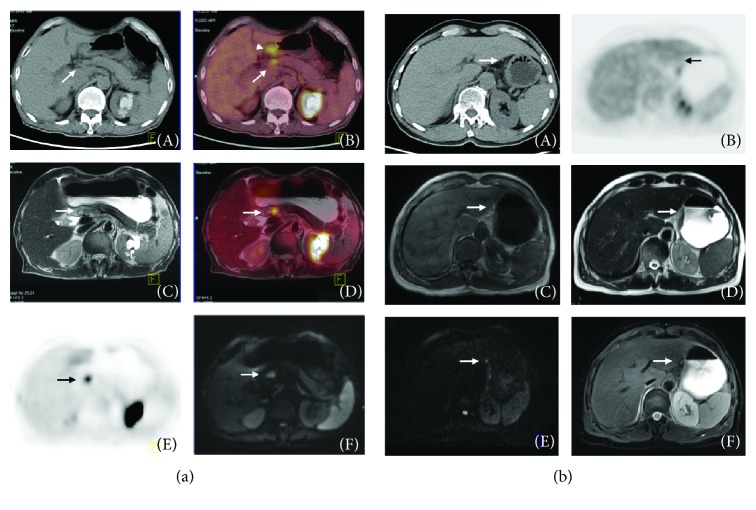
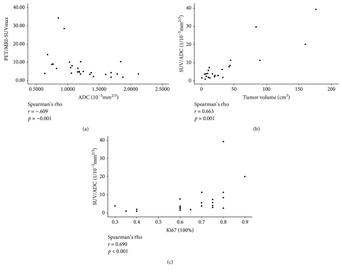
18F-FDG PET/MRI has been applied to the diagnosis and preoperative staging in various tumor types; however, reports using PET/MRI in gastric cancer are rare because of motion artifacts. We investigated the value of PET/MRI for preoperative staging compared with PET/CT in gastric cancer (GC). Thirty patients with confirmed GC underwent PET/CT and PET/MRI. TNM staging for each patient was determined from the PET/MRI and PET/CT images. The diagnostic performance of PET/MRI and PET/CT was calculated compared with the pathologic TNM stage. The two methods were compared using statistical analyses. The accuracy for T staging between PET/MRI and PET/CT was 76.9% vs. 57.7%, respectively. In T and T staging, the sensitivity and specificity for PET/MRI vs. PET/CT was 1.0 vs. 0.6 and 1.0 vs. 0.8, respectively. The area under the curve (AUC) for PET/MRI vs. PET/CT was 1.00 vs. 0.78 in the T stage, 0.73 vs. 0.66 in the T stage, 0.72 vs. 0.57 in the T stage, and 0.86 vs. 0.83 in the T stage. The accuracy for N staging of PET/MRI vs. PET/CT was 53.9% vs. 34.0%, and that for N vs. N was 85.0% vs. 77.0%. The sensitivity for PET/MRI in N3 staging was 0.67 and 0 for PET/CT. There was a statistically significant difference in the AUC for N staging (PET/MRI vs. PET/CT, 0.63 vs. 0.53, = 0.03). SUVmax/ADC positively correlated with tumor volume and Ki-67. PET/MRI performs more accurately in TNM staging compared with PET/CT and is optimal for accurate N staging. SUVmax/ADC has positive correlations with tumor volume and Ki-67.
18F-氟代脱氧葡萄糖正电子发射断层显像/磁共振成像(18F-FDG PET/MRI)已应用于多种肿瘤类型的诊断及术前分期;然而,由于运动伪影,关于PET/MRI在胃癌中的应用报道较少。我们研究了PET/MRI与PET/CT相比在胃癌(GC)术前分期中的价值。30例确诊为GC的患者接受了PET/CT和PET/MRI检查。根据PET/MRI和PET/CT图像确定每位患者的TNM分期。将PET/MRI和PET/CT的诊断性能与病理TNM分期进行比较计算。采用统计分析对这两种方法进行比较。PET/MRI和PET/CT在T分期的准确率分别为76.9%和57.7%。在T1和T2分期中,PET/MRI与PET/CT相比的敏感性和特异性分别为1.0对0.6和1.0对0.8。在T1期,PET/MRI与PET/CT的曲线下面积(AUC)分别为1.00和0.78;在T2期分别为0.73和0.66;在T3期分别为0.72和0.57;在T4期分别为0.86和0.83。PET/MRI与PET/CT在N分期的准确率分别为53.9%和34.0%,在N1与N2分期中分别为85.0%和77.0%。PET/MRI在N3分期的敏感性为0.67,PET/CT为0。在N分期的AUC方面存在统计学显著差异(PET/MRI对PET/CT,0.63对0.53,P = 0.03)。最大标准摄取值/表观扩散系数(SUVmax/ADC)与肿瘤体积和Ki-67呈正相关。与PET/CT相比,PET/MRI在TNM分期中表现更准确,且在准确的N分期方面最为理想。SUVmax/ADC与肿瘤体积和Ki-67呈正相关。