Laboratory of Neurobiology, School of Biomedical Engineering, Tianjin Medical University, Tianjin, China.
VCANBIO biological Resources Storage (Tianjin) Co. Ltd, Tianjin, China.
Sci Rep. 2019 Mar 14;9(1):4518. doi: 10.1038/s41598-019-41241-x.
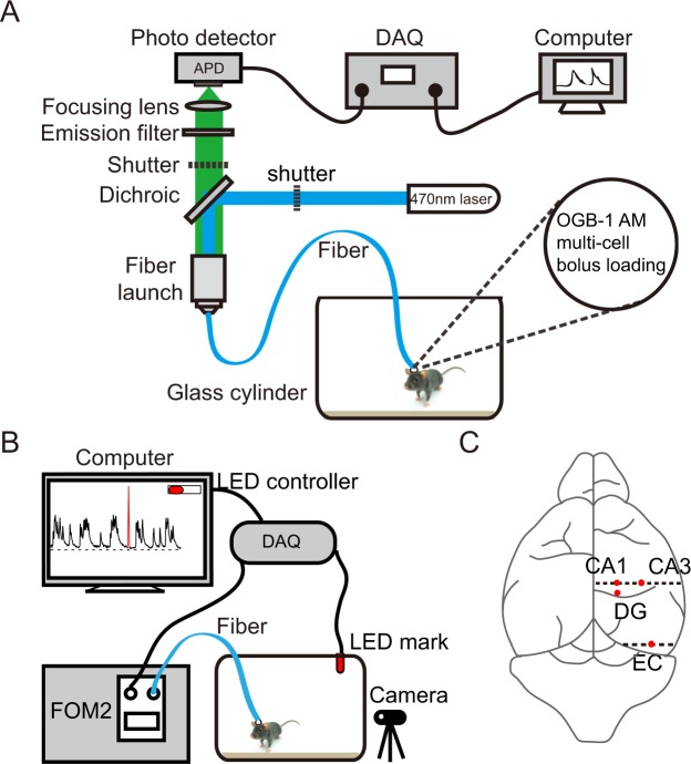
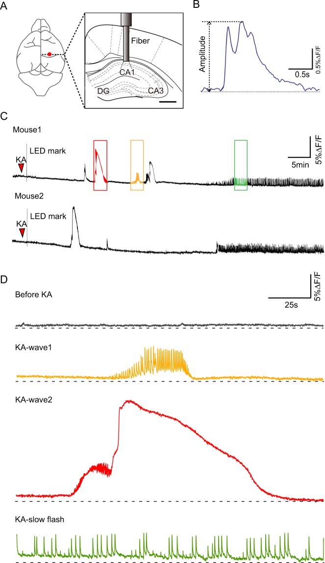
Epilepsy is a multi-etiological brain dysfunction syndrome. Hippocampal neuronal damage induced by seizures may be one of the causes leading to cognitive impairment, but the underlying mechanism remains to be further elucidated. The kainic acid (KA) model of temporal lobe epilepsy is widely used in understanding of the epileptogenesis. Fiber photometry is a signal detection technology suitable for recording calcium activity of neurons in the deep brain of freely moving animal. Here, we used the optical fiber-based method to monitor the real-time neuronal population activities of freely moving mice after subcutaneous injection of KA. We observed that KA administration led to one to three kinds of stereotypical patterns of epileptiform calcium activity in CA1, CA3, and dentate gyrus (DG) of the hippocampus, as well as the entorhinal cortex (EC). There were three kinds of waves in the hippocampal CA1, which we named wave 1, wave 2 and slow flash. Wave 1 and wave 2 appeared in both the CA3 and DG regions, but the EC only showed wave 1. In these epileptiform calcium signals, we observed a high amplitude and long duration calcium wave as a part of wave 2, which resembled cortical spreading depression (CSD) and always appeared at or after the end of seizure. Because the same characteristic of epileptiform calcium signal appeared in different brain regions, calcium signal may not exist with region specificity, but may exhibit a cell type specific manner. Thus, our work provides a support for the pathogenesis of epilepsy and epileptiform signal transmission research.
癫痫是一种多病因的脑功能障碍综合征。癫痫发作引起的海马神经元损伤可能是导致认知功能障碍的原因之一,但具体机制仍有待进一步阐明。颞叶癫痫的海人酸(KA)模型广泛应用于癫痫发生机制的研究。光纤光度测定法是一种适合记录自由活动动物深部脑神经元钙活性的信号检测技术。在这里,我们使用基于光纤的方法来监测 KA 皮下注射后自由活动小鼠海马 CA1、CA3 和齿状回(DG)以及内嗅皮层(EC)中神经元群体的实时活动。我们观察到 KA 给药导致海马 CA1、CA3 和 DG 以及内嗅皮层(EC)中出现一到三种癫痫样钙活性的刻板模式。在海马 CA1 中有三种波,我们分别命名为波 1、波 2 和慢闪光。波 1 和波 2 出现在 CA3 和 DG 区域,但 EC 仅显示波 1。在这些癫痫样钙信号中,我们观察到作为波 2 一部分的高振幅和长持续时间钙波,类似于皮质扩散性抑制(CSD),并且总是出现在发作结束时或之后。由于不同脑区出现相同特征的癫痫样钙信号,钙信号可能不存在区域特异性,而可能表现出细胞类型特异性方式。因此,我们的工作为癫痫发病机制和癫痫样信号传递研究提供了支持。