Eye Institute, Eye & ENT Hospital, Shanghai Medical College, Fudan University, Shanghai, China,
NHC Key Laboratory of Myopia, Fudan University, Key Laboratory of Myopia, Chinese Academy of Medical Sciences, Shanghai, China,
Int J Nanomedicine. 2019 Feb 25;14:1489-1501. doi: 10.2147/IJN.S195504. eCollection 2019.
Antiangiogenic drugs usually have short-acting efficacy and poor treatment compliance. The purpose of this study was to determine whether mesoporous silica nanoparticles (MSNs) could be utilized as a nanodrug delivery system for improving antiangiogenic therapy.
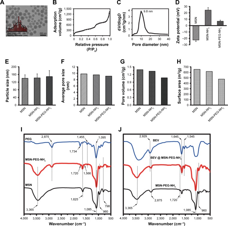
MSN-encapsulated bevacizumab nanoparticles were prepared by the nanocasting strategy and characterized by Fourier transform infrared, transmission electron microscopy, and Brunauer-Emmett-Teller method. Encapsulation efficiency and drug loading efficiency of MSN-encapsulated bevacizumab nanoparticles were calculated. The pharmacokinetics, cytotoxicity, and tissue toxicity were evaluated in vitro and in vivo. The antiangiogenic effects of MSN-bevacizumab nanoparticles were evaluated in vitro and in vivo.
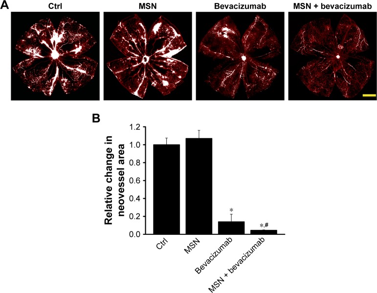
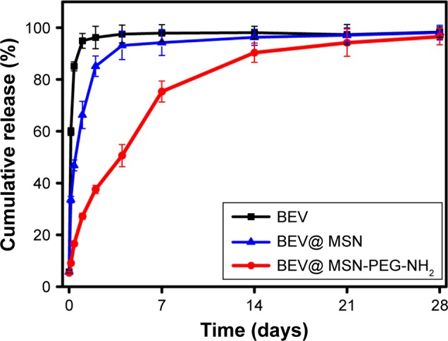
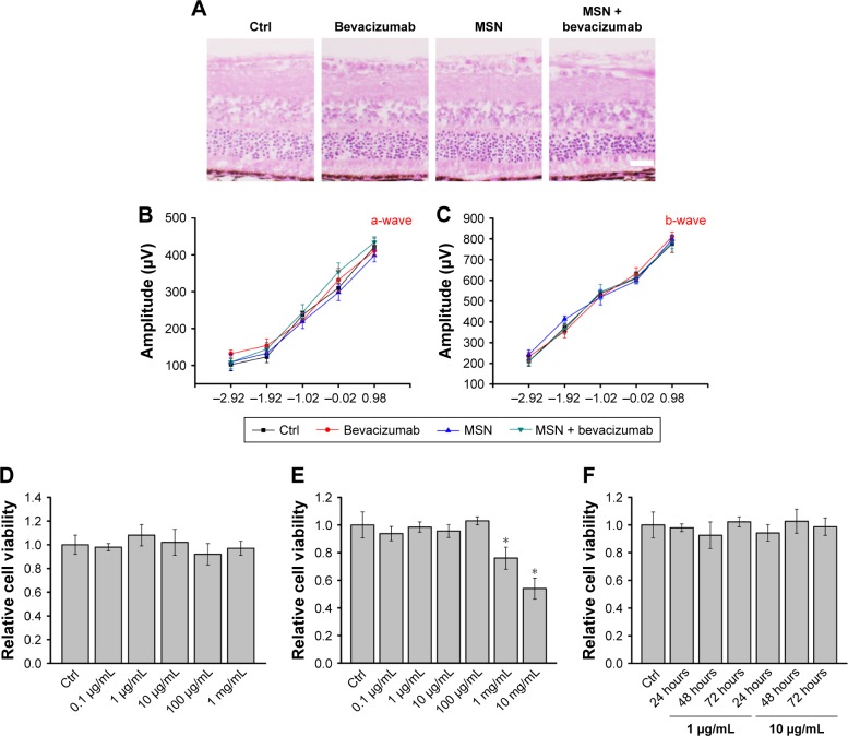
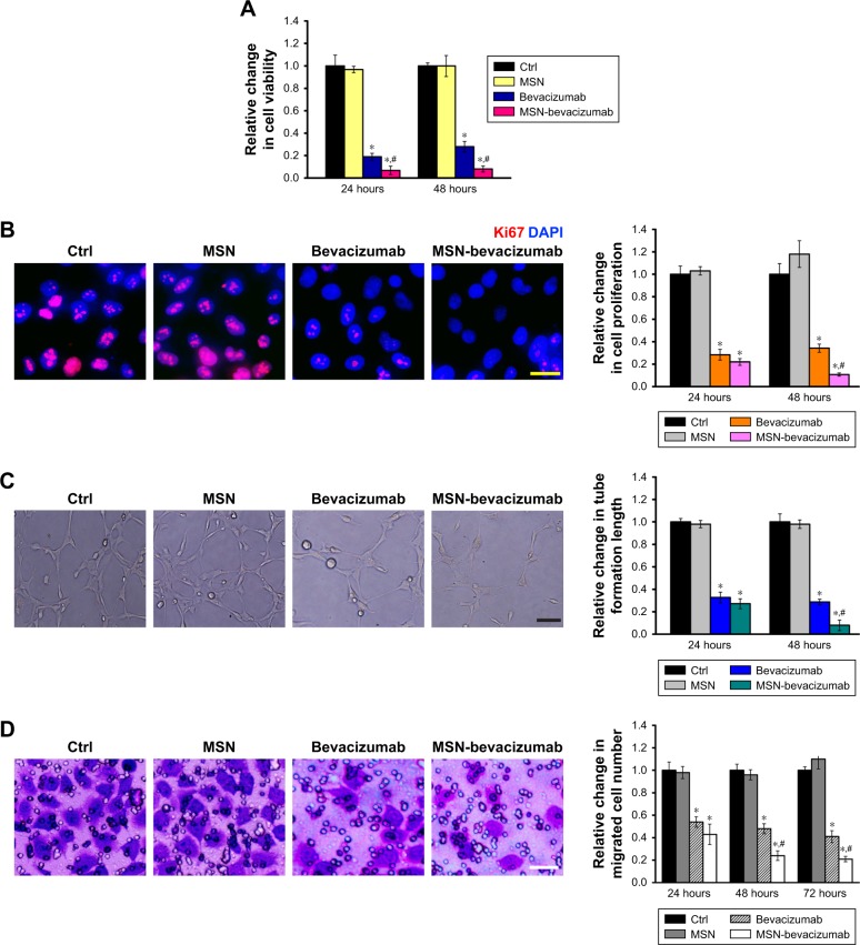
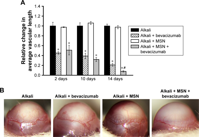
MSN encapsulation could prolong the residency of bevacizumab in vitreous/aqueous humor and maintain the long-lasting drug concentration. MSN-encapsulated bevacizumab nanoparticles did not show any obvious cytotoxicity and tissue toxicity. MSN-encapsulated bevacizumab nanoparticles were more effective than bevacizumab in suppressing vascular endothelial growth factor-induced endothelial cell proliferation, migration, and tube formation in vitro. MSN-encapsulated bevacizumab nanoparticles showed sustained inhibitory effects on corneal neovascularization and retinal neovascularization in vivo.
This study provides a novel strategy of encapsulating bevacizumab to protect and deliver it, which could increase the time between administration and formulation shelf-life. MSN-encapsulated bevacizumab is a promising drug delivery alternative of antiangiogenic therapy.
抗血管生成药物通常具有作用时间短和治疗顺应性差的特点。本研究旨在确定介孔硅纳米粒子(MSNs)是否可被用作改善抗血管生成治疗的纳米药物递送系统。
采用纳米铸型策略制备 MSN 包封贝伐珠单抗纳米粒,并通过傅里叶变换红外光谱、透射电子显微镜和 Brunauer-Emmett-Teller 法对其进行表征。计算 MSN 包封贝伐珠单抗纳米粒的包封效率和载药效率。在体外和体内评估其药代动力学、细胞毒性和组织毒性。在体外和体内评估 MSN-贝伐珠单抗纳米粒的抗血管生成作用。
MSN 包封可延长贝伐珠单抗在玻璃体内/房水中的滞留时间并维持长时间的药物浓度。MSN 包封的贝伐珠单抗纳米粒没有显示出明显的细胞毒性和组织毒性。MSN 包封的贝伐珠单抗纳米粒在抑制血管内皮生长因子诱导的内皮细胞增殖、迁移和管腔形成方面比贝伐珠单抗更有效。MSN 包封的贝伐珠单抗纳米粒在体内对角膜新生血管和视网膜新生血管具有持续的抑制作用。
本研究提供了一种将贝伐珠单抗包封以进行保护和递送的新策略,可增加给药时间和制剂货架寿命之间的时间间隔。MSN 包封的贝伐珠单抗是一种很有前途的抗血管生成治疗的药物递送替代物。