Instituto Carlos Chagas, Fundação Oswaldo Cruz (Fiocruz), Curitiba, Brazil.
Centro de Desenvolvimento Tecnológico em Saúde (CDTS), Fundação Oswaldo Cruz, Rio de Janeiro, Brazil.
mSphere. 2019 Mar 20;4(2):e00080-19. doi: 10.1128/mSphere.00080-19.
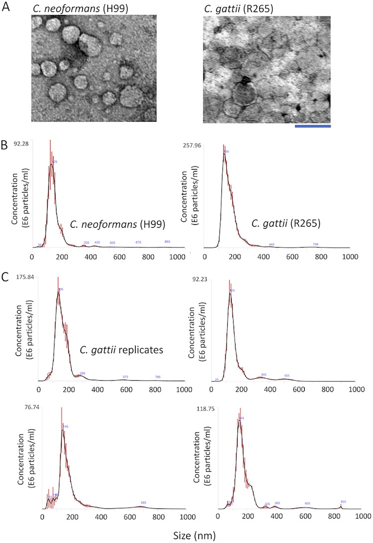
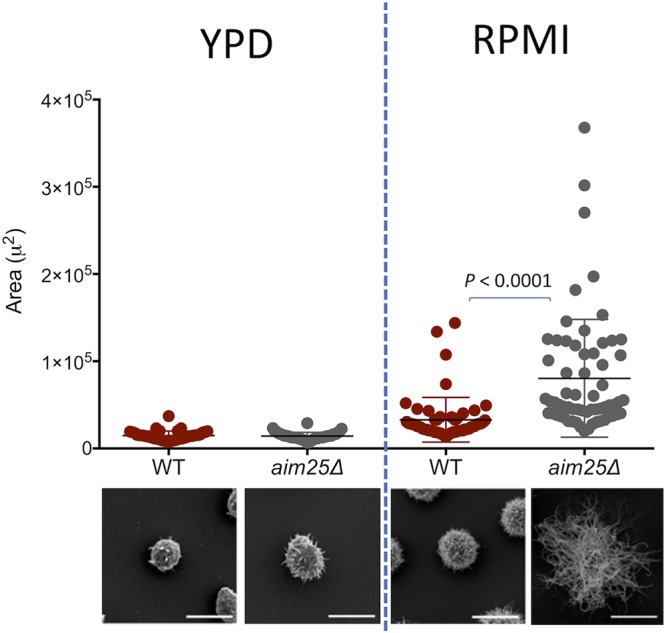
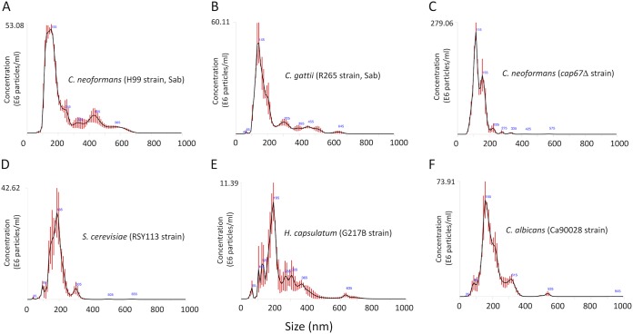
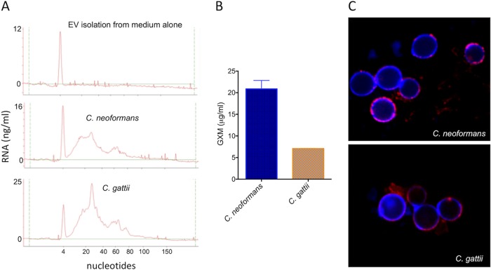
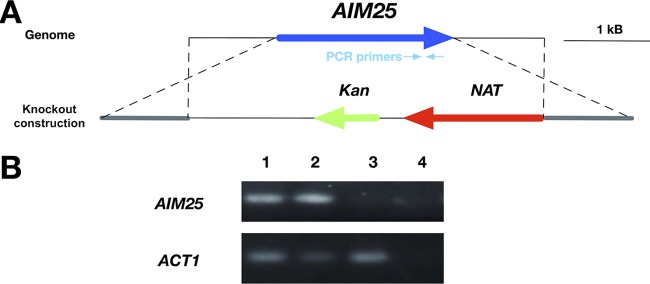
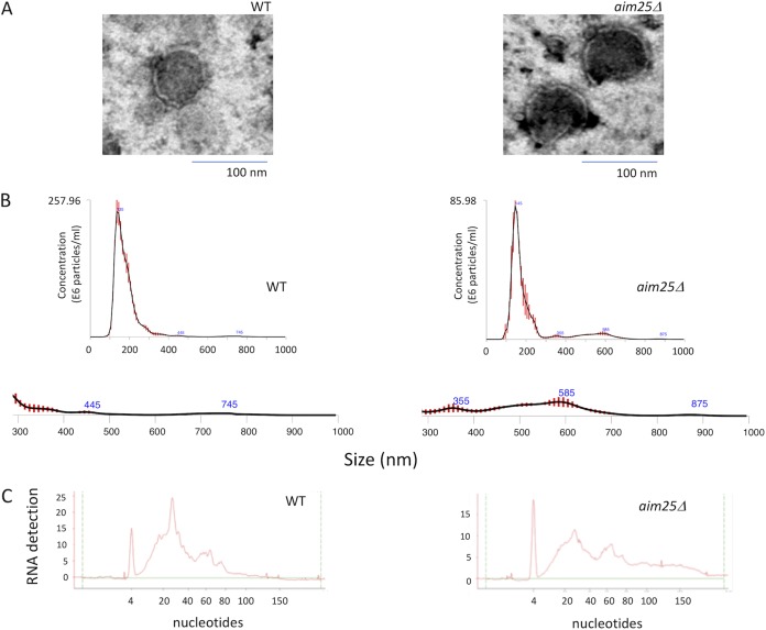
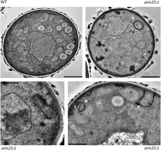
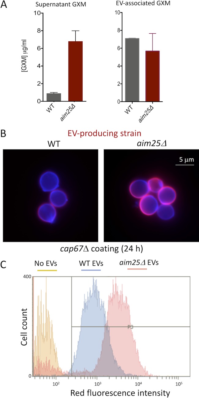
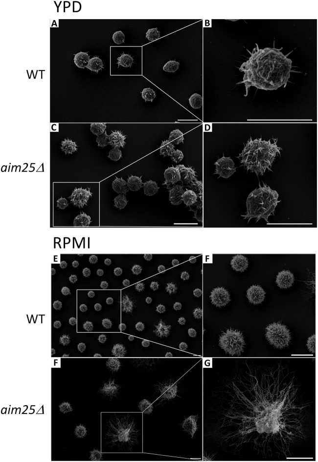
Regular protocols for the isolation of fungal extracellular vesicles (EVs) are time-consuming, hard to reproduce, and produce low yields. In an attempt to improve the protocols used for EV isolation, we explored a model of vesicle production after growth of and on solid media. Nanoparticle tracking analysis in combination with transmission electron microscopy revealed that and produced EVs in solid media. The properties of cryptococcal vesicles varied according to the culture medium used and the EV-producing species. EV detection was reproduced with an acapsular mutant of , as well as with isolates of , , and Cryptococcal EVs produced in solid media were biologically active and contained regular vesicular components, including the major polysaccharide glucuronoxylomannan (GXM) and RNA. Since the protocol had higher yields and was much faster than the regular methods used for the isolation of fungal EVs, we asked if it would be applicable to address fundamental questions related to cryptococcal secretion. On the basis that polysaccharide export in requires highly organized membrane traffic culminating with EV release, we analyzed the participation of a putative scramblase (Aim25; CNBG_3981) in EV-mediated GXM export and capsule formation in EVs from a Δ strain differed from those obtained from wild-type (WT) cells in physical-chemical properties and cargo. In a model of surface coating of an acapsular cryptococcal strain with vesicular GXM, EVs obtained from the Δ mutant were more efficiently used as a source of capsular polysaccharides. Lack of the Aim25 scramblase resulted in disorganized membranes and increased capsular dimensions. These results associate the description of a novel protocol for the isolation of fungal EVs with the identification of a previously unknown regulator of polysaccharide release. Extracellular vesicles (EVs) are fundamental components of the physiology of cells from all kingdoms. In pathogenic fungi, they participate in important mechanisms of transfer of antifungal resistance and virulence, as well as in immune stimulation and prion transmission. However, studies on the functions of fungal EVs are still limited by the lack of efficient methods for isolation of these compartments. In this study, we developed an alternative protocol for isolation of fungal EVs and demonstrated an application of this new methodology in the study of the physiology of the fungal pathogen Our results describe a fast and reliable method for the study of fungal EVs and reveal the participation of scramblase, a phospholipid-translocating enzyme, in secretory processes of .
常规的真菌细胞外囊泡(EVs)分离方法耗时、难以重现且产量低。为了改进 EV 分离的方法,我们探索了在固体培养基上生长后囊泡产生的模型。纳米颗粒跟踪分析与透射电子显微镜结合表明,和在固体培养基中产生 EV。隐球菌囊泡的特性根据使用的培养基和产生 EV 的物种而变化。用 的无荚膜突变体以及 和 的分离株重现了 EV 的检测, 在固体培养基中产生的 EV 具有生物活性并含有常规囊泡成分,包括主要多糖葡聚糖(GXM)和 RNA。由于该方案的产量更高且比用于真菌 EV 分离的常规方法快得多,我们想知道它是否适用于解决与隐球菌分泌相关的基本问题。基于 在 中多糖输出需要以 EV 释放为终点的高度有组织的膜运输,我们分析了假定的膜无序酶(Aim25;CNBG_3981)在 EV 介导的 GXM 输出和荚膜形成中的参与 在 Δ 株中的 EV 与从野生型(WT)细胞获得的 EV 在物理化学性质和货物方面有所不同。在无荚膜隐球菌菌株表面用囊泡 GXM 包被的模型中,从 Δ 突变体获得的 EV 更有效地用作荚膜多糖的来源。缺乏 Aim25 无序酶会导致膜无序和荚膜尺寸增加。这些结果将一种新型真菌 EV 分离方法的描述与一种以前未知的多糖释放调节剂的鉴定联系起来。细胞外囊泡(EVs)是所有生物界细胞生理学的基本组成部分。在致病性真菌中,它们参与了抗真菌耐药性和毒力的重要转移机制,以及免疫刺激和朊病毒传播。然而,由于缺乏分离这些隔室的有效方法,真菌 EVs 的功能研究仍然受到限制。在这项研究中,我们开发了一种分离真菌 EV 的替代方案,并证明了这种新方法在研究真菌病原体 的生理学中的应用。我们的结果描述了一种快速可靠的真菌 EV 研究方法,并揭示了无序酶(一种磷脂转运酶)在 的分泌过程中的参与。