Department of Obstetrics and Gynecology, Oakland University William Beaumont School of Medicine, Royal Oak, Michigan, United States of America.
Department of Obstetrics and Gynaecology, Wayne State University School of Medicine, Detroit, Michigan, United States of America.
PLoS One. 2019 Mar 21;14(3):e0200229. doi: 10.1371/journal.pone.0200229. eCollection 2019.
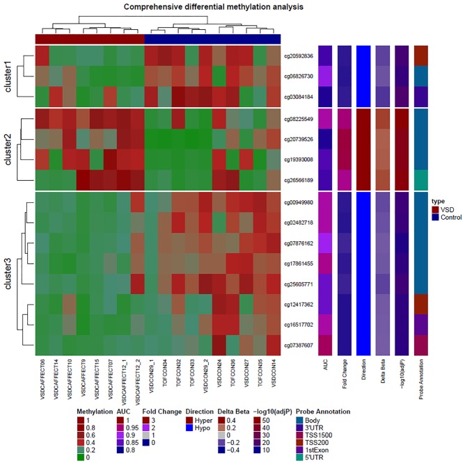
Ventricular Septal Defect (VSD), the most common congenital heart defect, is characterized by a hole in the septum between the right and left ventricles. The pathogenesis of VSD is unknown in most clinical cases. There is a paucity of data relevant to epigenetic changes in VSD. The placenta is a fetal tissue crucial in cardiac development and a potentially useful surrogate for evaluating the development of heart tissue. To understand epigenetic mechanisms that may play a role in the development of VSD, genome-wide DNA methylation assay on placentas of 8 term subjects with isolated VSD and no known or suspected genetic syndromes and 10 unaffected controls was performed using the Illumina HumanMethylation450 BeadChip assay. We identified a total of 80 highly accurate potential CpGs in 80 genes for detection of VSD; area under the receiver operating characteristic curve (AUC ROC) 1.0 with significant 95% CI (FDR) p-values < 0.05 for each individual locus. The biological processes and functions for many of these differentially methylated genes are previously known to be associated with heart development or disease, including cardiac ventricle development (HEY2, ISL1), heart looping (SRF), cardiac muscle cell differentiation (ACTC1, HEY2), cardiac septum development (ISL1), heart morphogenesis (SRF, HEY2, ISL1, HEYL), Notch signaling pathway (HEY2, HEYL), cardiac chamber development (ISL1), and cardiac muscle tissue development (ACTC1, ISL1). In addition, we identified 8 microRNAs that have the potential to be biomarkers for the detection of VSD including: miR-191, miR-548F1, miR-148A, miR-423, miR-92B, miR-611, miR-2110, and miR-548H4. To our knowledge this is the first report in which placental analysis has been used for determining the pathogenesis of and predicting VSD.
室间隔缺损(VSD)是最常见的先天性心脏病,其特征是左右心室之间的间隔有一个洞。大多数临床病例的 VSD 发病机制尚不清楚。关于 VSD 中表观遗传变化的数据很少。胎盘是胎儿心脏发育过程中至关重要的胎儿组织,是评估心脏组织发育的潜在有用替代物。为了了解可能在 VSD 发育中起作用的表观遗传机制,我们对 8 名患有孤立性 VSD 且无已知或可疑遗传综合征的足月受试者的胎盘和 10 名未受影响的对照者进行了全基因组 DNA 甲基化检测,使用了 Illumina HumanMethylation450 BeadChip 检测。我们总共鉴定了 80 个在 80 个基因中具有高度准确性的潜在 CpG 来检测 VSD;每个个体位点的接收者操作特征曲线(AUC ROC)下面积为 1.0,具有显著的 95%CI(FDR)p 值<0.05。这些差异甲基化基因的许多生物过程和功能先前与心脏发育或疾病有关,包括心室发育(HEY2、ISL1)、心脏环化(SRF)、心肌细胞分化(ACTC1、HEY2)、心脏间隔发育(ISL1)、心脏形态发生(SRF、HEY2、ISL1、HEYL)、Notch 信号通路(HEY2、HEYL)、心腔发育(ISL1)和心肌组织发育(ACTC1、ISL1)。此外,我们还鉴定了 8 个具有成为 VSD 检测生物标志物潜力的 microRNA,包括:miR-191、miR-548F1、miR-148A、miR-423、miR-92B、miR-611、miR-2110 和 miR-548H4。据我们所知,这是首次利用胎盘分析来确定 VSD 的发病机制和预测 VSD 的报告。