Jousi Mikko O, Erkkilä Jukka, Varjonen Mari, Soiva Martti, Hukkinen Katja, Blanco Sequeiros Roberto
Päijät-Häme Central Hospital, Lahti, Finland.
Planmed Oy, Helsinki, Finland.
Acta Radiol Open. 2019 Mar 15;8(3):2058460119836255. doi: 10.1177/2058460119836255. eCollection 2019 Mar.
Digital breast tomosynthesis (DBT) is gaining popularity in breast imaging. There are several different technical approaches for conducting DBT imaging.
To determine optimal imaging parameters, test patient friendliness, evaluate the initial diagnostic performance, and describe diagnostic advances possible with the new Continuous Sync-and-Shoot method.
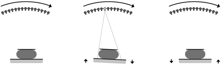
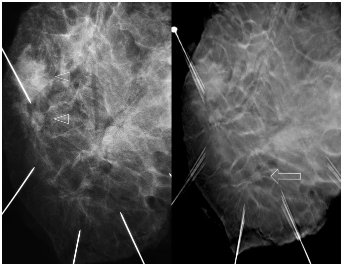
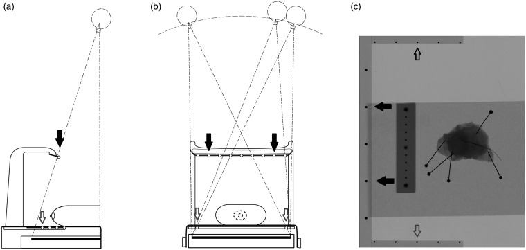
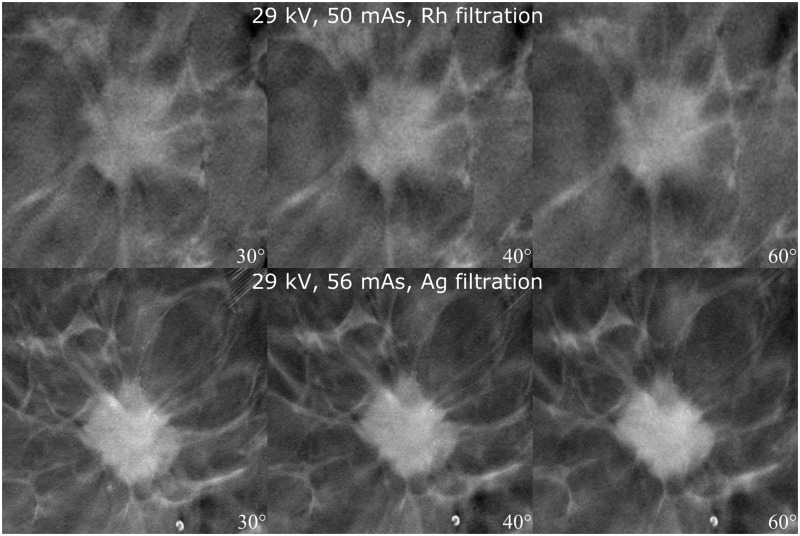
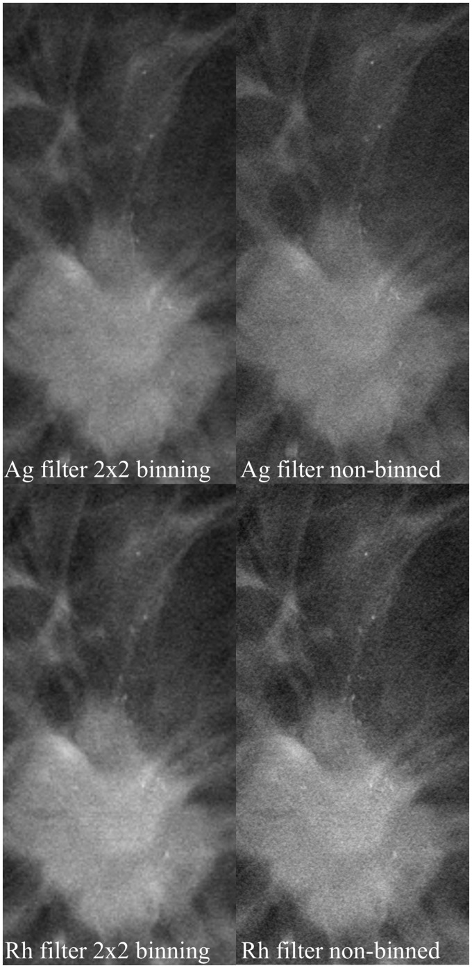
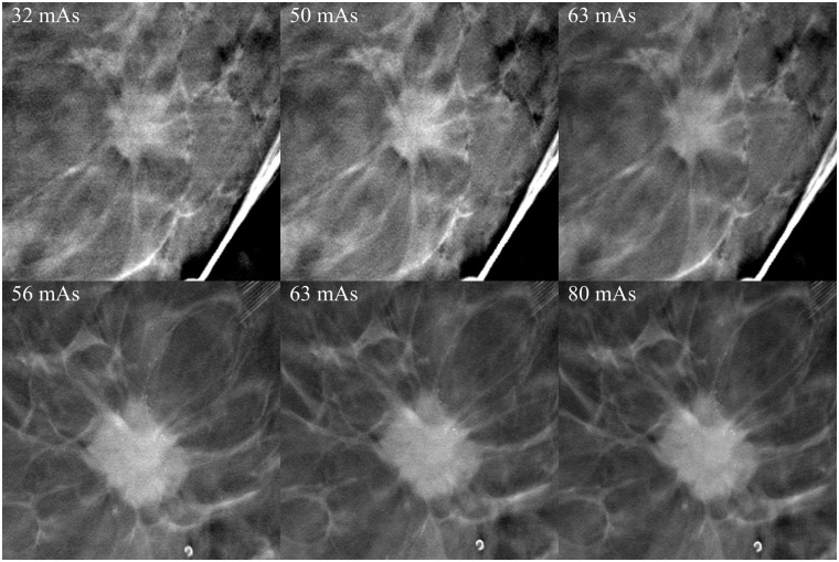
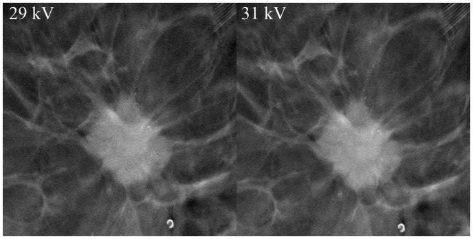
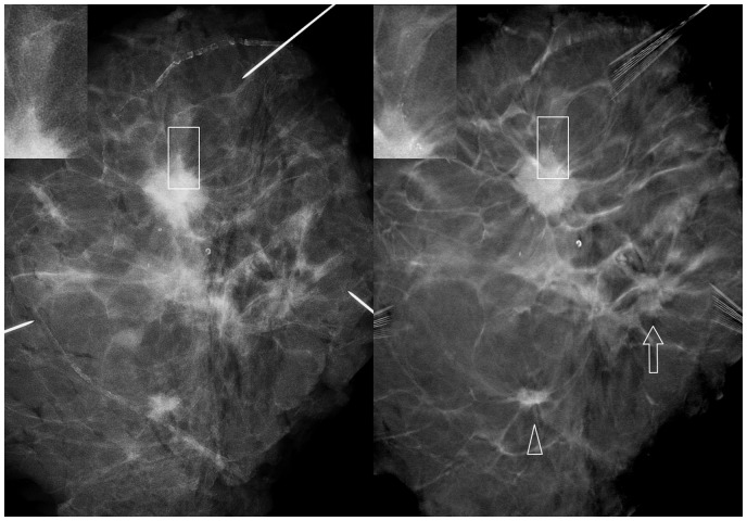
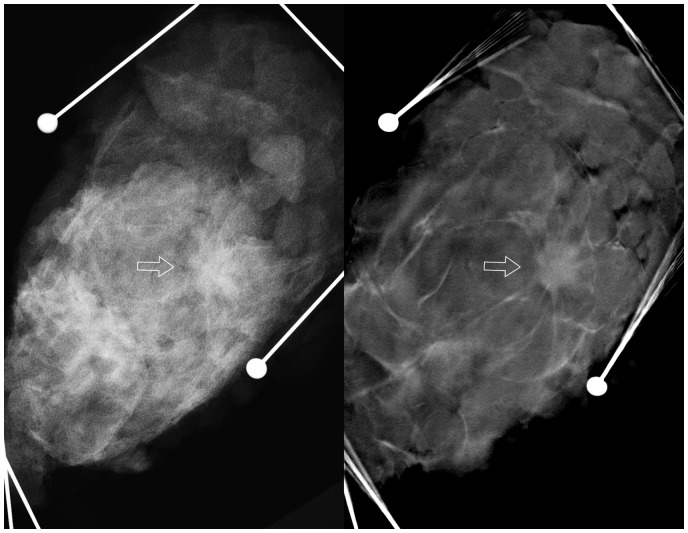
Thirty-six surgical breast specimens were imaged with digital mammography (DM) and a prototype of a DBT system (Planmed Oy, Helsinki, Finland). We tested the patient friendliness of the sync-and-shoot movement without radiation exposure in eight volunteers. Different imaging parameters were tested with 20 specimens to identify the optimal combination: angular range 30°, 40°, and 60°; pixel binning; Rhodium (Rh) and Silver (Ag) filtrations; and different kV and mAs values. Two breast radiologists evaluated 16 DM and DBT image pairs and rated six different image properties. Imaging modalities were compared with paired t-test.
The Continuous Sync-and-Shoot method produced diagnostically valid images. Five out of eight volunteers felt no/minimal discomfort, three experienced mild discomfort from the tilting movement of the detector, with the motion being barely recognized. The combination of 30°, Ag filtering, and 2 × 2 pixel binning produced the best image quality at an acceptable dose level. DBT was significantly better in all six evaluated properties ( < 0.05). Mean Dose/Dose ratio was 1.22 (SD = 0.42).
The evaluated imaging method is feasible for imaging and analysing surgical breast specimens and DBT is significantly better than DM in image evaluation.
数字乳腺断层合成(DBT)在乳腺成像中越来越受欢迎。进行DBT成像有几种不同的技术方法。
确定最佳成像参数,测试患者友好性,评估初始诊断性能,并描述采用新的连续同步拍摄方法可能实现的诊断进展。
用数字乳腺摄影(DM)和DBT系统原型(芬兰赫尔辛基的普兰梅德公司)对36个手术切除的乳腺标本进行成像。我们在8名志愿者中测试了无辐射暴露情况下同步拍摄运动的患者友好性。用20个标本测试不同的成像参数以确定最佳组合:角度范围30°、40°和60°;像素合并;铑(Rh)和银(Ag)滤过;以及不同的千伏和毫安秒值。两名乳腺放射科医生评估了16对DM和DBT图像,并对六种不同的图像特性进行评分。采用配对t检验比较成像方式。
连续同步拍摄方法产生了具有诊断价值的图像。8名志愿者中有5人感觉无不适/轻微不适,3人因探测器的倾斜运动感到轻微不适,这种运动几乎难以察觉。30°、Ag滤过和2×2像素合并的组合在可接受的剂量水平下产生了最佳图像质量。在所有六项评估特性中,DBT均显著更好(<0.05)。平均剂量/剂量比为1.22(标准差=0.42)。
所评估的成像方法对于手术切除的乳腺标本成像和分析是可行的,并且在图像评估方面DBT明显优于DM。