Department of Psychology University of Rome "Sapienza" Rome Italy.
Department of Biomedical and Neuromotor Sciences (DIBINEM) University of Bologna Bologna Italy.
Ann Clin Transl Neurol. 2019 Feb 1;6(3):445-455. doi: 10.1002/acn3.718. eCollection 2019 Mar.
Narcolepsy type 1 widely affects the architecture of sleep with frequent fast transition to REM sleep at both nighttime and daytime sleep onset. The occurrence of repeated sleep onset REM periods over the Multiple Sleep Latency Test offers a unique opportunity to identify EEG patterns predictive of successful dream recall after short periods composed of only REM or NREM sleep. It also permits to disentangle state- from trait-like differences in dream recall, by using a within-subjects design.
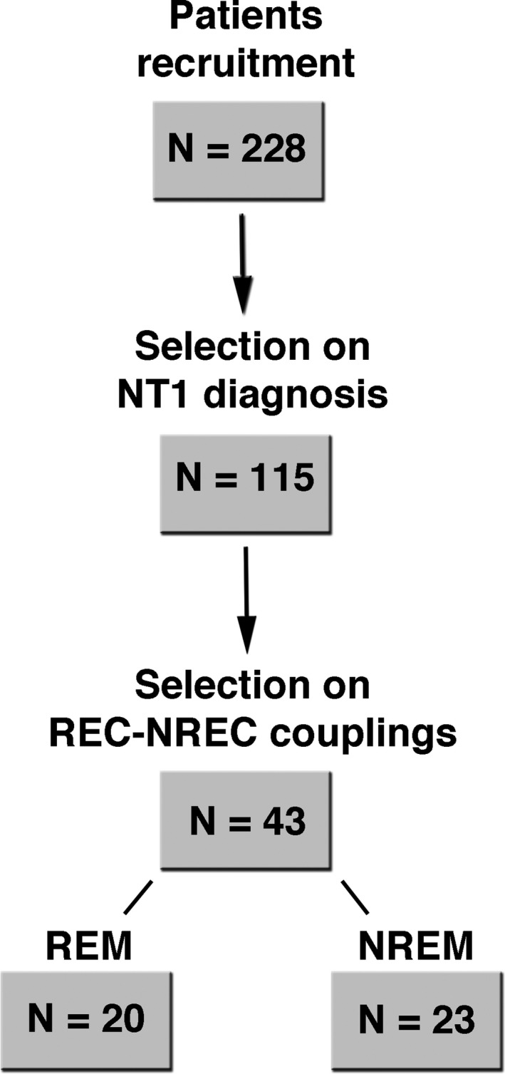
A consecutive series of 115 first-diagnosed drug-free adult narcolepsy-type 1 patients underwent Multiple Sleep Latency Tests and were asked after each nap opportunity if they had or had not a dream experience. Scalp EEG power and a specific index of cortical activation (delta/beta power ratio), obtained from naps of 43 patients with both presence and absence of dream recall in the same sleep stage, were compared separately for REM and NREM sleep.
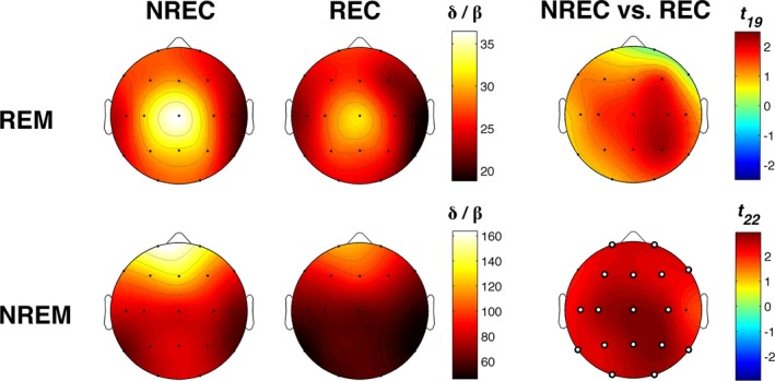
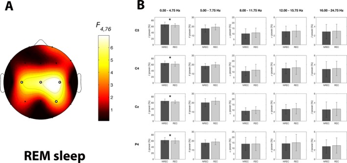
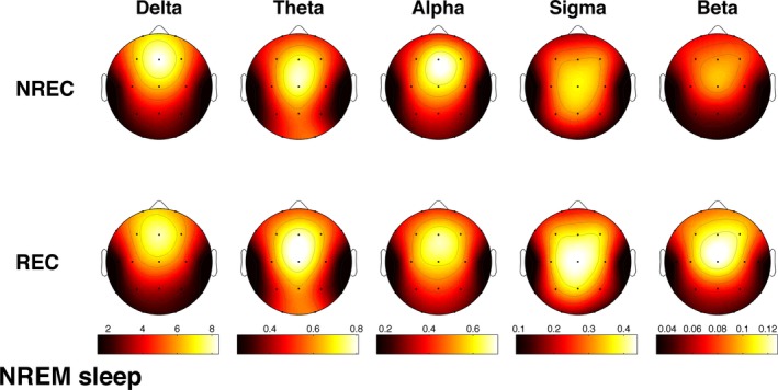
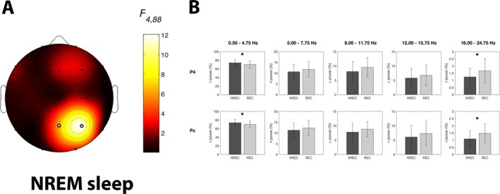
Successful dream recall was associated with an increased EEG desynchronization in both REM and NREM over partially overlapping cortical areas. Compared to unsuccessful recall, it showed (1) lower delta power over centro-parietal areas during both stages, (2) higher beta power in the same cortical areas during NREM, and (3) lower values in the delta/beta ratio during NREM in most scalp locations.
A more activated electrophysiological milieu in both REM and NREM sleep promotes dream recall, strengthening the notion that the parietal areas are crucial not only in generating dream experience, as shown in brain-damaged patients, but also in the memory processing leading to recall.
1 型发作性睡病广泛影响睡眠结构,导致夜间和白天睡眠起始时快速进入 REM 睡眠期。多次睡眠潜伏期试验(MSLT)中反复出现 REM 睡眠起始期,为识别对 REM 或 NREM 睡眠期极短时间后成功回忆梦境的脑电图(EEG)模式提供了独特的机会。该试验还允许使用受试者内设计,区分梦回忆中的状态与特质差异。
连续纳入 115 例未经药物治疗的首次确诊的成年 1 型发作性睡病患者,进行多次睡眠潜伏期试验,在每个小睡机会后询问他们是否有梦境体验。对 43 例患者 REM 和 NREM 睡眠期均有且无梦境回忆的小睡进行分析,比较头皮 EEG 功率和皮质激活的特定指标(δ/β 功率比)。
成功的梦境回忆与 REM 和 NREM 睡眠中部分重叠的皮质区域的 EEG 去同步化增加有关。与不成功的回忆相比,其表现为:(1)在两个阶段中,中顶区的 δ 波功率降低;(2)NREM 时相同皮质区域的 β 波功率升高;(3)大多数头皮位置的 NREM 时 δ/β 比值降低。
在 REM 和 NREM 睡眠中,更活跃的电生理环境促进了梦境回忆,这进一步证明了顶区不仅在生成梦境体验中很重要,正如在脑损伤患者中所显示的那样,而且在导致回忆的记忆处理中也很重要。