Chang Chen-Ming, Lee Brian J, Grant Alexander M, Groll Andrew N, Levin Craig S
Departments of Applied Physics and Radiology, Stanford University, Stanford, CA, USA.
Departments of Mechanical Engineering and Radiology, Stanford University, Stanford, CA, USA.
IEEE Trans Radiat Plasma Med Sci. 2018 Sep;2(5):422-431. doi: 10.1109/TRPMS.2018.2852686. Epub 2018 Jul 3.
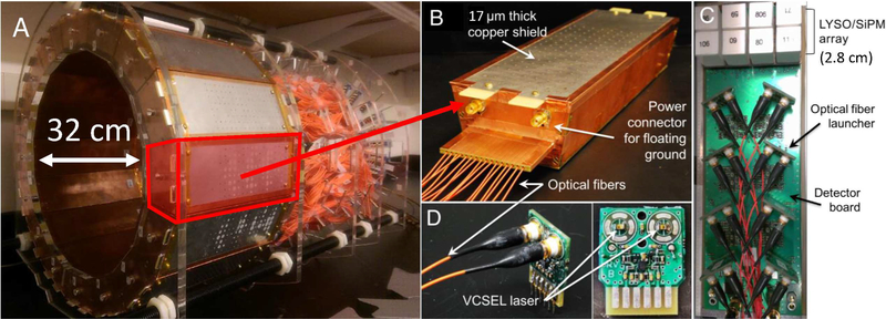
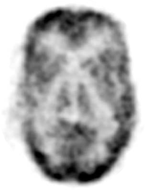
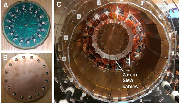
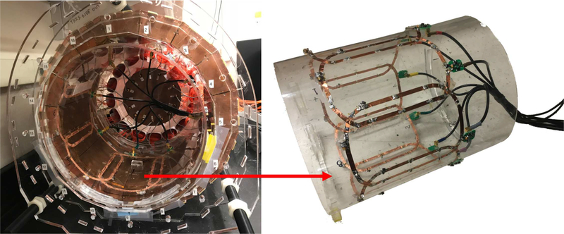
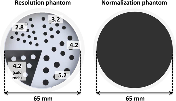
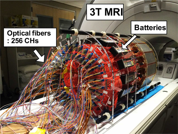
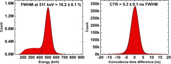
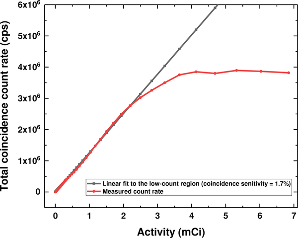
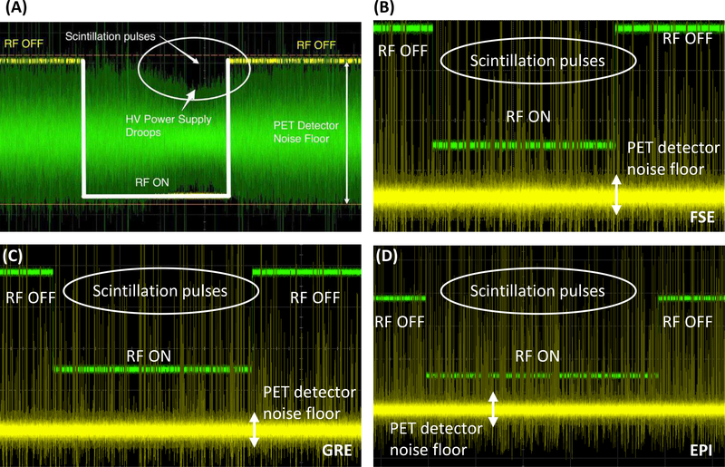
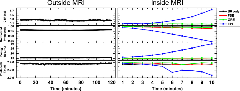
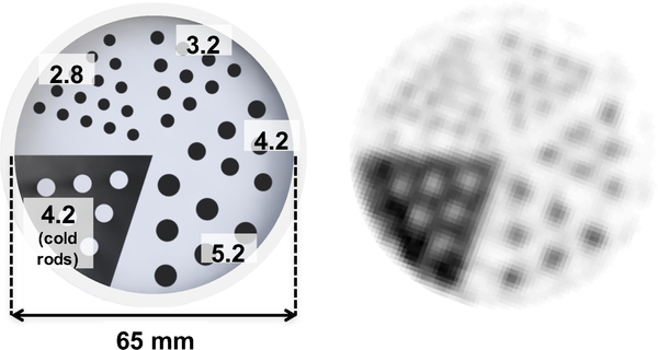
Hybrid positron emission tomography (PET)/magnetic resonance imaging (MRI) has risen to the cutting edge of medical imaging technology as it allows simultaneous acquisition of structural, functional and molecular information of the patient. A PET insert that can be installed into existing MR systems can in principle reduce the cost barriers for an existing MR site to achieve simultaneous PET/MRI compared to procuring an integrated PET+MRI system. The PET insert systems developed so far for PET/MRI require the RF transmitter coil to reside inside the PET ring as those PET inserts block the RF fields from the MRI system. Here we report for the first time on the performance of a full-ring brain-sized "RF-penetrable" PET insert we have recently completed. This insert allows the RF fields generated by the built-in body coil to penetrate the PET ring. The PET insert comprises a ring of 16 detector modules employing electro-optical coupled signal transmission and a multiplexing framework based on compressed sensing. Energy resolution, coincidence timing resolution (CTR), photopeak position, and coincidence count rate were acquired outside and inside a 3-Tesla MRI system under simultaneous acquisition to evaluate the impact of MRI on the PET performance. Coincidence count rate performance was evaluated by acquiring a cylinder source with high initial activity decaying over time. Tomographic imaging of two phantoms, a custom 6.5-cm diameter resolution phantom with hot rods of four different sizes (2.8 mm, 3.2 mm, 4.2 mm, and 5.2 mm diameter) and a 3D Hoffman brain phantom, were performed to evaluate the imaging capability of the PET insert. The energy resolution at 511 keV and CTR acquired by the PET insert were 16.2±0.1% and 5.3±0.1 ns FWHM, respectively, and remained stable during MRI operation except when the EPI sequence was applied. The PET system starts to show saturation in coincidence count rate at 2.76 million photon counts per second. Most of the 2.8-mm diameter hot rods and main features of the 3D Hoffman brain phantom were resolved by the PET insert, demonstrating its high spatial resolution and capability to image a complex tracer distribution mimicking that seen in the human brain.
混合正电子发射断层扫描(PET)/磁共振成像(MRI)已跃升至医学成像技术的前沿,因为它能够同时获取患者的结构、功能和分子信息。与采购集成式PET+MRI系统相比,一种可安装到现有MR系统中的PET插入件原则上可以降低现有MR站点实现同步PET/MRI的成本障碍。到目前为止,为PET/MRI开发的PET插入件系统要求射频发射线圈位于PET环内,因为这些PET插入件会阻挡来自MRI系统的射频场。在此,我们首次报告了我们最近完成的全环脑尺寸“射频可穿透”PET插入件的性能。该插入件允许内置体线圈产生的射频场穿透PET环。该PET插入件包括一个由16个探测器模块组成的环,采用电光耦合信号传输以及基于压缩感知的复用框架。在3特斯拉MRI系统内外同时采集时,获取了能量分辨率、符合定时分辨率(CTR)、光电峰位置和符合计数率,以评估MRI对PET性能的影响。通过采集一个初始活度较高且随时间衰减的圆柱源来评估符合计数率性能。对两个体模进行断层成像,一个是定制的直径6.5厘米的分辨率体模,带有四种不同尺寸(直径2.8毫米、3.2毫米、4.2毫米和5.2毫米)的热棒,另一个是3D霍夫曼脑体模,以评估PET插入件的成像能力。PET插入件在511keV处的能量分辨率和获取的CTR分别为16.2±0.1%和5.3±0.1纳秒半高宽,并且在MRI运行期间保持稳定,除了应用回波平面成像(EPI)序列时。PET系统在每秒276万个光子计数时开始显示符合计数率饱和。PET插入件分辨出了大多数直径2.8毫米的热棒以及3D霍夫曼脑体模的主要特征,证明了其高空间分辨率以及对模拟人脑所见复杂示踪剂分布进行成像的能力。