Tissue Engineering Group (TEG), Department of Orthopaedic Surgery (NOCERAL), Faculty of Medicine, University of Malaya, Kuala Lumpur, Malaysia.
Musculoskeletal Research Group, Department of Molecular and Clinical Cancer Medicine, Institute of Translational Medicine, Liverpool, United Kingdom.
PLoS One. 2019 Mar 27;14(3):e0214212. doi: 10.1371/journal.pone.0214212. eCollection 2019.
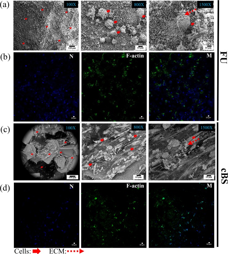
It has been demonstrated that nanocrystalline forsterite powder synthesised using urea as a fuel in sol-gel combustion method had produced a pure forsterite (FU) and possessed superior bioactive characteristics such as bone apatite formation and antibacterial properties. In the present study, 3D-scaffold was fabricated using nanocrystalline forsterite powder in polymer sponge method. The FU scaffold was used in investigating the physicochemical, biomechanics, cell attachment, in vitro biocompatibility and osteogenic differentiation properties. For physicochemical characterisation, Fourier-transform infrared spectroscopy (FTIR), Energy dispersive X-ray (EDX), X-ray diffraction (XRD), Raman spectroscopy, X-ray photoemission spectrometer (XPS) and Brunauer-Emmett-Teller (BET) were used. FTIR, EDX, XRD peaks and Raman spectroscopy demonstrated correlating to FU. The XPS confirmed the surface chemistry associating to FU. The BET revealed FU scaffold surface area of 12.67 m2/g and total pore size of 0.03 cm3/g. Compressive strength of the FU scaffold was found to be 27.18 ± 13.4 MPa. The human bone marrow derived mesenchymal stromal cells (hBMSCs) characterisation prior to perform seeding on FU scaffold verified the stromal cell phenotypic and lineage commitments. SEM, confocal images and presto blue viability assay suggested good cell attachment and proliferation of hBMSCs on FU scaffold and comparable to a commercial bone substitutes (cBS). Osteogenic proteins and gene expression from day 7 onward indicated FU scaffold had a significant osteogenic potential (p<0.05), when compared with day 1 as well as between FU and cBS. These findings suggest that FU scaffold has a greater potential for use in orthopaedic and/or orthodontic applications.
已经证明,使用尿素作为燃料在溶胶-凝胶燃烧法中合成的纳米晶镁橄榄石粉末产生了纯镁橄榄石(FU),并且具有优异的生物活性特性,如骨磷灰石形成和抗菌性能。在本研究中,使用纳米晶镁橄榄石粉末通过聚合物海绵法制备了 3D 支架。FU 支架用于研究物理化学、生物力学、细胞附着、体外生物相容性和成骨分化特性。进行物理化学特性分析时,使用了傅里叶变换红外光谱(FTIR)、能量色散 X 射线(EDX)、X 射线衍射(XRD)、拉曼光谱、X 射线光电子能谱(XPS)和 Brunauer-Emmett-Teller(BET)。FTIR、EDX、XRD 峰和拉曼光谱证明与 FU 相关。XPS 证实了与 FU 相关的表面化学。BET 显示 FU 支架的表面积为 12.67 m2/g,总孔体积为 0.03 cm3/g。FU 支架的抗压强度为 27.18±13.4 MPa。在将 hBMSCs 接种到 FU 支架上之前,对人骨髓间充质基质细胞(hBMSCs)进行了特性鉴定,验证了基质细胞表型和谱系承诺。SEM、共聚焦图像和 presto blue 活力测定表明,hBMSCs 在 FU 支架上具有良好的细胞附着和增殖能力,与商业骨替代物(cBS)相当。从第 7 天开始的成骨蛋白和基因表达表明,与第 1 天相比,FU 支架具有显著的成骨潜力(p<0.05),并且与 FU 和 cBS 之间也有显著差异。这些发现表明,FU 支架在骨科和/或正畸应用中具有更大的潜力。