Chen Hao, Xu Baofeng, Wang Guangming, Guo Yunbao, Hou Kun, Yu Jinlu
Department of Neurosurgery, The First Hospital of Jilin University, Changchun, China.
Medicine (Baltimore). 2019 Mar;98(13):e14678. doi: 10.1097/MD.0000000000014678.
Intermuscular hemangioma (IH) usually occurs in the muscles of the limbs and trunk, but can rarely occur in the occipital region. IH in the occipital region is easily misdiagnosed as arteriovenous malformation (AVM).
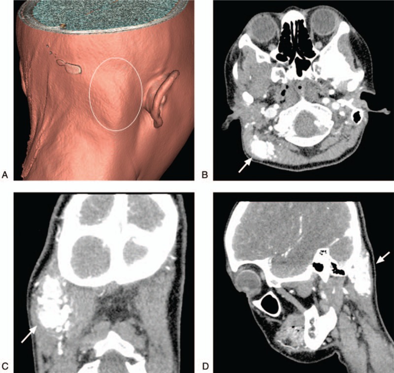
A 31-year-old woman had a right occipital mass for 5 months without pulsation.
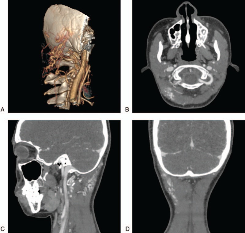
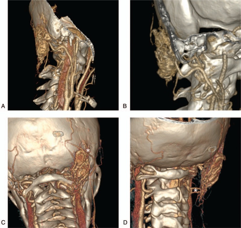
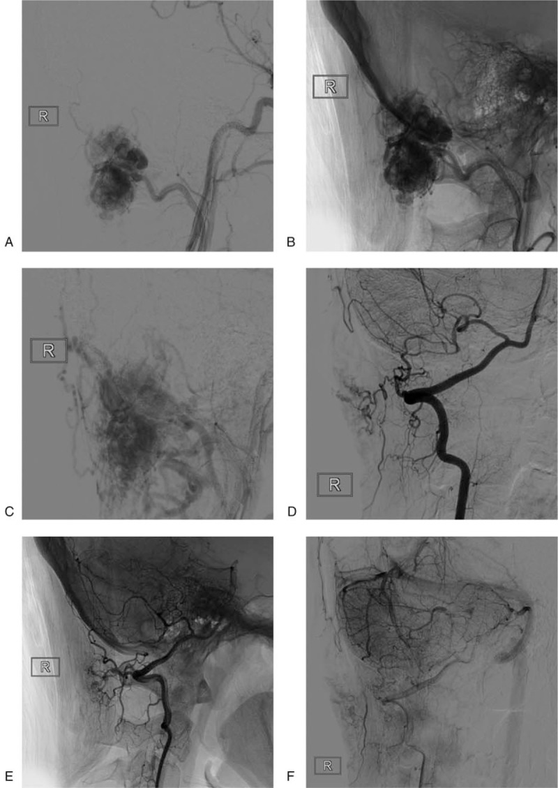
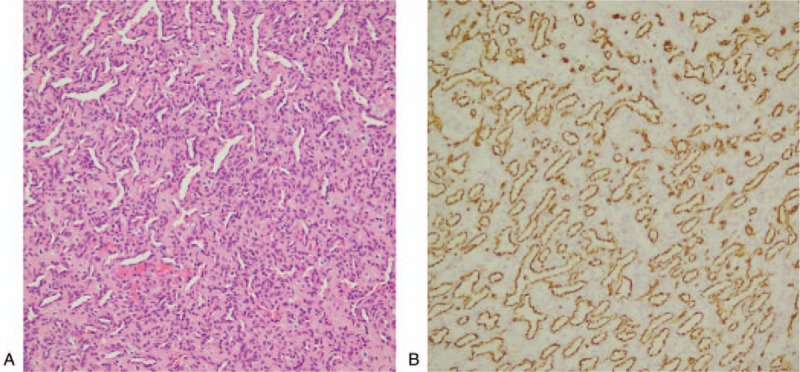
Head computered tomography angiography (CTA) and digital substraction angiography (DSA) examinations showed that the lesion was mainly vascular, approximately 3 × 5 cm in size, and supplied by occipital arteries and the muscular branches of vertebral arteries. The venous drainage of the lesions communicated with the suboccipital vein plexus and the paravertebral vein in the venous phase, indicating AVM. Postoperative histological investigation showed the lesion was a hemangioma.
It was recommended for surgical removal. The surgery was carried out under general anesthesia. The lesion showed a clear boundary. The occipital artery touched the anterior margin of the lesion, was exposed and ligated, and was removed around the lesion. The lesion consisted of massive blood vessels, and the surrounding muscles were swollen, indicating IH.After the lesion was removed, the normal muscle tissue around the lesion was also removed.
The patient achieved a good recovery after surgery, and pathology confirmed IH. A postoperative 1-year CTA review was performed and showed partial residual, then the radiotherapy was recommended. She refused further radiotherapy, follow-up 2 years later showed no enlargement of the lesion.
Although IH rarely occurs in the occipital region, this can occur. Due to the complexity of the drainage veins in the occipital region, these IH are prone to misdiagnosis as AVM.
肌间血管瘤(IH)通常发生于四肢和躯干的肌肉,但很少发生于枕部区域。枕部区域的IH易被误诊为动静脉畸形(AVM)。
一名31岁女性右枕部肿物5个月,无搏动。
头部计算机断层血管造影(CTA)和数字减影血管造影(DSA)检查显示,病变主要为血管性,大小约3×5 cm,由枕动脉和椎动脉肌支供血。病变的静脉引流在静脉期与枕下静脉丛和椎旁静脉相通,提示为AVM。术后组织学检查显示病变为血管瘤。
建议手术切除。在全身麻醉下进行手术。病变边界清晰。枕动脉触及病变前缘,予以暴露并结扎,沿病变周围切除。病变由大量血管组成,周围肌肉肿胀,提示为IH。病变切除后,病变周围的正常肌肉组织也被切除。
患者术后恢复良好,病理证实为IH。术后1年进行CTA复查显示有部分残留,遂建议放疗。患者拒绝进一步放疗,2年后随访显示病变未增大。
尽管IH很少发生于枕部区域,但仍有可能发生。由于枕部区域引流静脉的复杂性,这些IH容易被误诊为AVM。