West China School of Medicine, West China Hospital, Sichuan University, No. 37 Guoxue Alley, Chengdu 610041, China.
State Key Laboratory of Biotherapy and Cancer Center, West China Hospital, Sichuan University, Chengdu 610041, China.
Contrast Media Mol Imaging. 2019 Feb 25;2019:4507694. doi: 10.1155/2019/4507694. eCollection 2019.
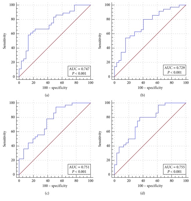
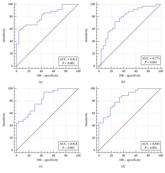
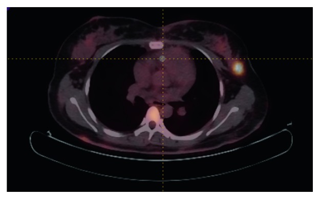
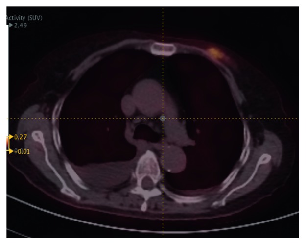
. To investigate the value of SUV metrics and radiomic features based on the ability of F-FDG PET/CT in differentiating between breast lymphoma and breast carcinoma. . A total of 67 breast nodules from 44 patients who underwent F-FDG PET/CT pretreatment were retrospectively analyzed. Radiomic parameters and SUV metrics were extracted using the LIFEx package on PET and CT images. All texture parameters were divided into six groups: histogram (HISTO), SHAPE, gray-level co-occurrence matrix (GLCM), gray-level run-length matrix (GLRLM), neighborhood gray-level different matrix (NGLDM), and gray-level zone-length matrix (GLZLM). Receiver operating characteristics (ROC) curves were generated to evaluate the discriminative ability of each parameter, and the optimal parameter in each group was selected to generate a new predictive variable by using binary logistic regression. PET predictive variable, CT predictive variable, the combination of PET and CT predictive variables, and SUV were compared in terms of areas under the curve (AUCs), sensitivity, specificity, and accuracy. . Except for SUVmin (=0.971), the averages of FDG uptake metrics of lymphoma were significantly higher than those of carcinoma ( ≤ 0.001), with the following median values: SUV, 4.75 versus 2.38 g/ml ( < 0.001); SUV, 2.04 versus 0.88 g/ml (=0.001); SUV, 10.69 versus 4.76 g/ml (=0.001); SUV, 9.15 versus 2.78 g/ml ( < 0.001); TLG, 42.24 versus 9.90 ( < 0.001). In the ROC curves analysis based on radiomic features and SUV, the AUC for SUV was 0.747, for CT texture parameters was 0.729, for PET texture parameters was 0.751, and for the combination of CT and PET texture parameters was 0.771. . The SUV metrics in FDG PET/CT images showed a potential ability in the differentiation between breast lymphoma and carcinoma. The combination of SUV and PET/CT texture analysis may be promising to provide an effectively discriminant modality for the differential diagnosis of breast lymphoma and carcinoma, even for the differentiation of subtypes of lymphoma.
. 旨在研究基于 F-FDG PET/CT 区分乳腺淋巴瘤和乳腺癌的能力的 SUV 指标和放射组学特征的价值。. 回顾性分析了 44 例接受 F-FDG PET/CT 预处理的患者的 67 个乳腺结节。使用 PET 和 CT 图像上的 LIFEx 包提取放射组学参数和 SUV 指标。所有纹理参数分为六组:直方图(HISTO)、形状(SHAPE)、灰度共生矩阵(GLCM)、灰度游程长度矩阵(GLRLM)、邻域灰度差异矩阵(NGLDM)和灰度区域长度矩阵(GLZLM)。生成受试者工作特征(ROC)曲线以评估每个参数的区分能力,并通过二元逻辑回归选择每组中的最佳参数来生成新的预测变量。比较了 PET 预测变量、CT 预测变量、PET 和 CT 预测变量的组合以及 SUV 在曲线下面积(AUC)、敏感性、特异性和准确性方面的表现。. 除 SUVmin(=0.971)外,淋巴瘤的 FDG 摄取指标平均值明显高于乳腺癌(≤0.001),中位数如下:SUV,4.75 与 2.38g/ml(<0.001);SUV,2.04 与 0.88g/ml(=0.001);SUV,10.69 与 4.76g/ml(<0.001);SUV,9.15 与 2.78g/ml(<0.001);TLG,42.24 与 9.90(<0.001)。在基于放射组学特征和 SUV 的 ROC 曲线分析中,SUV 的 AUC 为 0.747,CT 纹理参数为 0.729,PET 纹理参数为 0.751,CT 和 PET 纹理参数的组合为 0.771。. FDG PET/CT 图像中的 SUV 指标在区分乳腺淋巴瘤和乳腺癌方面具有潜在能力。SUV 与 PET/CT 纹理分析的结合可能有望为乳腺淋巴瘤和乳腺癌的鉴别诊断提供一种有效的鉴别方式,甚至可以区分淋巴瘤的亚型。