Department of Biochemistry , Duke University School of Medicine , Durham , North Carolina 27710 , United States.
Department of Chemistry , Indian Institute of Science Education and Research Bhopal , Bhopal 462066 , India.
Biochemistry. 2019 Apr 16;58(15):1963-1974. doi: 10.1021/acs.biochem.9b00027. Epub 2019 Apr 5.
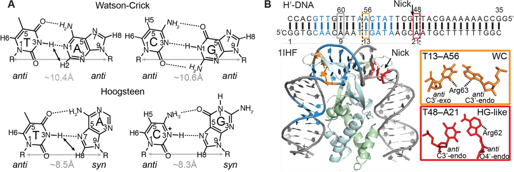
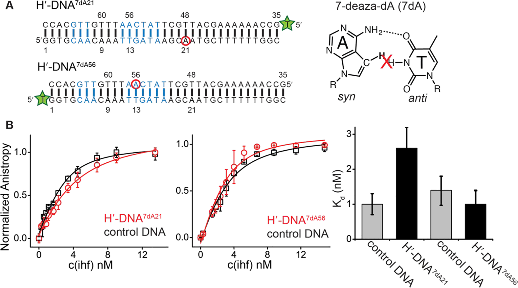
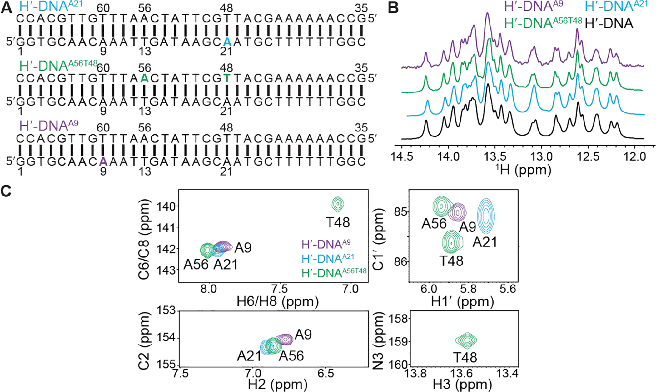
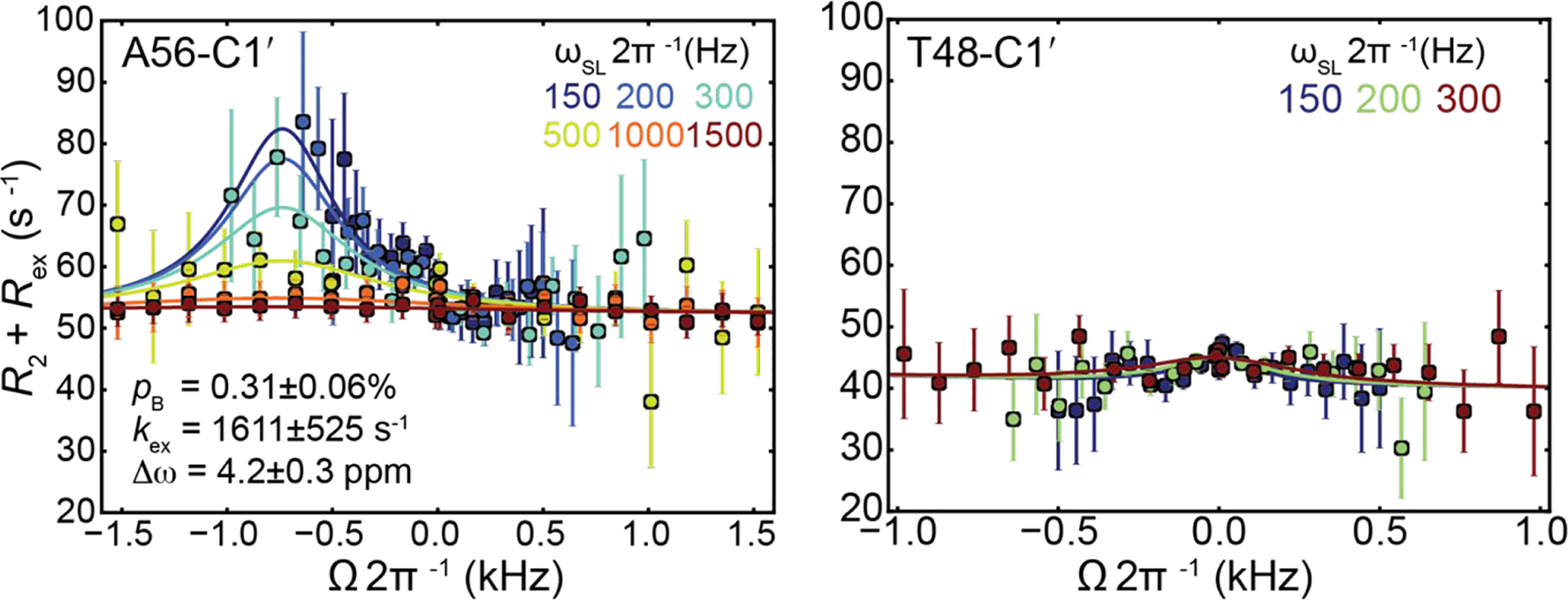
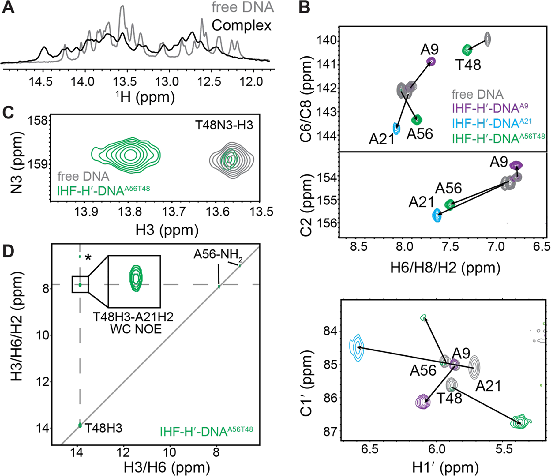
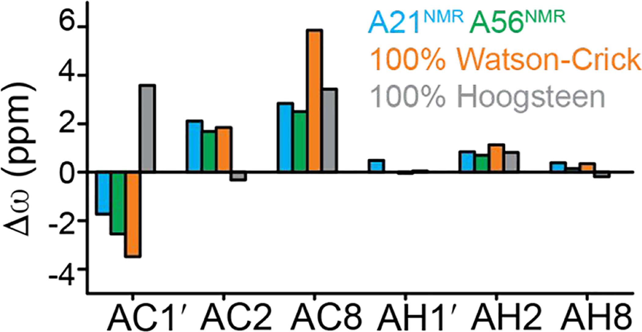
A( syn)-T and G( syn)-C Hoogsteen base pairs in protein-bound DNA duplexes can be difficult to resolve by X-ray crystallography due to ambiguous electron density and by nuclear magnetic resonance (NMR) spectroscopy due to poor chemical shift dispersion and size limitations with solution-state NMR spectroscopy. Here we describe an NMR strategy for characterizing Hoogsteen base pairs in protein-DNA complexes, which relies on site-specifically incorporating C- and N-labeled nucleotides into DNA duplexes for unambiguous resonance assignment and to improve spectral resolution. The approach was used to resolve the conformation of an A-T base pair in a crystal structure of an ∼43 kDa complex between a 34 bp duplex DNA and the integration host factor (IHF) protein. In the crystal structure (Protein Data Bank entry 1IHF ), this base pair adopts an unusual Hoogsteen conformation with a distorted sugar backbone that is accommodated by a nearby nick used to aid in crystallization. The NMR chemical shifts and interproton nuclear Overhauser effects indicate that this base pair predominantly adopts a Watson-Crick conformation in the intact DNA-IHF complex under solution conditions. Consistent with these NMR findings, substitution of 7-deazaadenine at this base pair resulted in only a small (∼2-fold) decrease in the IHF-DNA binding affinity. The NMR strategy provides a new approach for resolving crystallographic ambiguity and more generally for studying the structure and dynamics of protein-DNA complexes in solution.
在蛋白质结合的 DNA 双链中,A(顺)-T 和 G(顺)-C 霍加斯顿碱基对由于电子密度不明确,通过 X 射线晶体学难以解析,并且由于核磁共 振(NMR)光谱的化学位移分散性差和溶液 NMR 光谱的尺寸限制,通过 NMR 光谱也难以解析。在这里,我们描述了一种用于表征蛋白质-DNA 复合物中霍加斯顿碱基对的 NMR 策略,该策略依赖于将 C 和 N 标记的核苷酸特异性掺入 DNA 双链中,以进行明确的共振分配,并提高光谱分辨率。该方法用于解析约 43 kDa 大小的 34 个碱基对双链 DNA 与整合宿主因子(IHF)蛋白之间的复合物的晶体结构中 A-T 碱基对的构象。在晶体结构(蛋白数据银行条目 1IHF)中,该碱基对采用不寻常的霍加斯顿构象,糖骨架扭曲,由附近的缺口容纳,该缺口用于辅助结晶。NMR 化学位移和质子间核 Overhauser 效应表明,在完整的 DNA-IHF 复合物中,该碱基对在溶液条件下主要采用 Watson-Crick 构象。与这些 NMR 发现一致,在该碱基对处取代 7-脱氮腺嘌呤仅导致 IHF-DNA 结合亲和力略有下降(约 2 倍)。该 NMR 策略为解析晶体学中的歧义提供了一种新方法,并且更普遍地为研究溶液中蛋白质-DNA 复合物的结构和动力学提供了一种新方法。