Takaki Tadashi, Inagaki Azusa, Chonabayashi Kazuhisa, Inoue Keiji, Miki Kenji, Ohno Seiko, Makiyama Takeru, Horie Minoru, Yoshida Yoshinori
Department of Cell Growth and Differentiation, Center for iPS Cell Research and Application, Kyoto University, Sakyo-ku, Kyoto 606-8507, Japan.
Department of Cardiology, Japanese Red Cross Kyoto Daini Hospital, Kamigyo-ku, Kyoto 602-8026, Japan.
Stem Cells Int. 2019 Mar 6;2019:7532657. doi: 10.1155/2019/7532657. eCollection 2019.
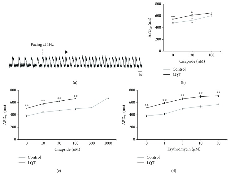
Induced pluripotent stem cells (iPSCs) from type 1 long QT (LQT1) patients can differentiate into cardiomyocytes (CMs) including ventricular cells to recapitulate the disease phenotype. Although optical recordings using membrane potential dyes to monitor action potentials (APs) were reported, no study has investigated the disease phenotypes of cardiac channelopathy in association with the cardiac subtype at the single-cell level. We induced iPSC-CMs from three control and three LQT1 patients. Single-cell analysis using a fast-responding dye confirmed that ventricular cells were the dominant subtype (control-iPSC-CMs: 98%, 88%, 91%; LQT1-iPSC-CMs: 95%, 79%, 92%). In addition, LQT1-iPSC-ventricular cells displayed an increased frequency of early afterdepolarizations (value = 0.031). Cardiomyocyte monolayers constituted mostly of ventricular cells derived from LQT1-iPSCs showed prolonged AP duration (APD) (value = 0.000096). High-throughput assays using cardiomyocyte monolayers in 96-well plates demonstrated that I inhibitors prolonged APDs in both control- and LQT1-iPSC-CM monolayers. We confirmed that the optical recordings of APs in single cells and monolayers derived from control- and LQT1-iPSC-CMs can be used to assess arrhythmogenicity, supporting the feasibility of membrane potential dye-based high-throughput screening to study ventricular arrhythmias caused by genetic channelopathy or cardiotoxic drugs.
1型长QT综合征(LQT1)患者的诱导多能干细胞(iPSC)可分化为心肌细胞(CM),包括心室细胞,以重现疾病表型。尽管已有报道使用膜电位染料进行光学记录来监测动作电位(AP),但尚无研究在单细胞水平上研究与心脏亚型相关的心脏离子通道病的疾病表型。我们从三名对照者和三名LQT1患者中诱导生成了iPSC-CM。使用快速响应染料进行的单细胞分析证实,心室细胞是主要亚型(对照-iPSC-CM:98%、88%、91%;LQT1-iPSC-CM:95%、79%、92%)。此外,LQT1-iPSC-心室细胞的早期后去极化频率增加(值=0.031)。由源自LQT1-iPSC的心室细胞构成的心肌细胞单层显示动作电位时程(APD)延长(值=0.000096)。在96孔板中使用心肌细胞单层进行的高通量检测表明,I类抑制剂可延长对照和LQT1-iPSC-CM单层的APD。我们证实,对照和LQT1-iPSC-CM来源的单细胞和单层细胞的AP光学记录可用于评估致心律失常性,支持基于膜电位染料的高通量筛选用于研究由遗传离子通道病或心脏毒性药物引起的室性心律失常的可行性。