Lima Zeinab Safarpour, Ebadi Mohammad Reza, Amjad Ghazaleh, Younesi Ladan
Shahid Akbarabadi Clinical Research Development Unit (ShACRDU), Iran University of Medical Sciences (IUMS), Tehran, Iran.
Shohadaye Haft-e-tir Hospital, Iran University of Medical Sciences (IUMS), Tehran, Iran.
Open Access Maced J Med Sci. 2019 Mar 14;7(5):838-848. doi: 10.3889/oamjms.2019.171. eCollection 2019 Mar 15.
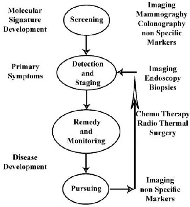
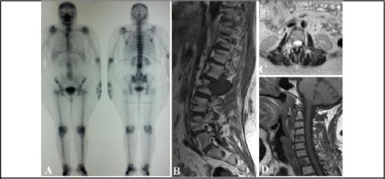
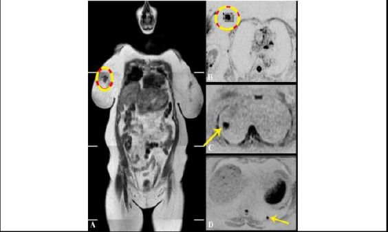
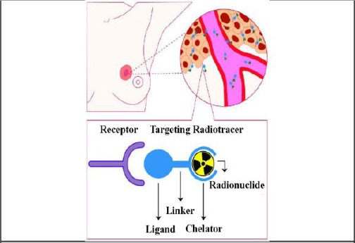
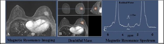
One of the techniques utilised in the management of cancer in all stages is multiple biomedical imaging. Imaging as an important part of cancer clinical protocols can provide a variety of information about morphology, structure, metabolism and functions. Application of imaging technics together with other investigative apparatus including in fluids analysis and vitro tissue would help clinical decision-making. Mixed imaging techniques can provide supplementary information used to improve staging and therapy planning. Imaging aimed to find minimally invasive therapy to make better results and reduce side effects. Probably, the most important factor in reducing mortality of certain cancers is an early diagnosis of cancer via screening based on imaging. The most common cancer in women is breast cancer. It is considered as the second major cause of cancer deaths in females, and therefore it remained as an important medical and socio-economic issue. Medical imaging has always formed part of breast cancer care and has used in all phases of cancer management from detection and staging to therapy monitoring and post-therapeutic follow-up. An essential action to be performed in the preoperative staging of breast cancer based on breast imaging. The general term of breast imaging refers to breast sonography, mammography, and magnetic resonance tomography (MRT) of the breast (magnetic resonance mammography, MRM). Further development in technology will lead to increase imaging speed to meet physiological processes requirements. One of the issues in the diagnosis of breast cancer is sensitivity limitation. To overcome this limitation, complementary imaging examinations are utilised that traditionally includes screening ultrasound, and combined mammography and ultrasound. Development in targeted imaging and therapeutic agents calls for close cooperation among academic environment and industries such as biotechnological, IT and pharmaceutical industries.
在癌症各阶段管理中使用的技术之一是多种生物医学成像。成像作为癌症临床方案的重要组成部分,可以提供有关形态、结构、代谢和功能的各种信息。将成像技术与包括体液分析和体外组织分析在内的其他检查设备一起应用,有助于临床决策。混合成像技术可以提供用于改善分期和治疗计划的补充信息。成像旨在寻找微创治疗方法,以取得更好的效果并减少副作用。也许,降低某些癌症死亡率的最重要因素是通过基于成像的筛查早期诊断癌症。女性中最常见的癌症是乳腺癌。它被认为是女性癌症死亡的第二大主要原因,因此仍然是一个重要的医学和社会经济问题。医学成像一直是乳腺癌护理的一部分,并已用于癌症管理的各个阶段,从检测、分期到治疗监测和治疗后随访。基于乳腺成像在乳腺癌术前分期中必须采取的一项重要行动。乳腺成像的一般术语是指乳腺超声、乳腺X线摄影和乳腺磁共振断层扫描(MRT)(磁共振乳腺摄影,MRM)。技术的进一步发展将提高成像速度,以满足生理过程的要求。乳腺癌诊断中的一个问题是灵敏度限制。为了克服这一限制,采用了传统上包括筛查超声以及乳腺X线摄影和超声联合检查在内的补充成像检查。靶向成像和治疗剂的发展需要学术环境与生物技术、信息技术和制药等行业密切合作。