• 文献检索
  • 文档翻译
  • 深度研究
  • 学术资讯
  • Suppr Zotero 插件Zotero 插件
  • 邀请有礼
  • 套餐&价格
  • 历史记录
应用&插件
Suppr Zotero 插件Zotero 插件浏览器插件Mac 客户端Windows 客户端微信小程序
定价
高级版会员购买积分包购买API积分包
服务
文献检索文档翻译深度研究API 文档MCP 服务
关于我们
关于 Suppr公司介绍联系我们用户协议隐私条款
关注我们

Suppr 超能文献

核心技术专利:CN118964589B侵权必究
粤ICP备2023148730 号-1Suppr @ 2026

文献检索

告别复杂PubMed语法,用中文像聊天一样搜索,搜遍4000万医学文献。AI智能推荐,让科研检索更轻松。

立即免费搜索

文件翻译

保留排版,准确专业,支持PDF/Word/PPT等文件格式,支持 12+语言互译。

免费翻译文档

深度研究

AI帮你快速写综述,25分钟生成高质量综述,智能提取关键信息,辅助科研写作。

立即免费体验

基于人工智能 CAD 的回顾性对比研究:降低乳腺 X 线摄影中的假阳性标记

Reduction of False-Positive Markings on Mammograms: a Retrospective Comparison Study Using an Artificial Intelligence-Based CAD.

机构信息

Department of Diagnostic Imaging, Breast Imaging Division, MD Anderson Center, University of Texas, 1515 Holcombe Blvd., Houston, TX, 77030, USA.

Voxel Imaging, Inc., 2711 N. Sepulveda Blvd., #284, Manhattan Beach, CA, 90266, USA.

出版信息

J Digit Imaging. 2019 Aug;32(4):618-624. doi: 10.1007/s10278-018-0168-6.

DOI:10.1007/s10278-018-0168-6
PMID:30963339
原文链接:https://pmc.ncbi.nlm.nih.gov/articles/PMC6646646/
Abstract

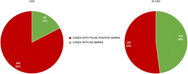
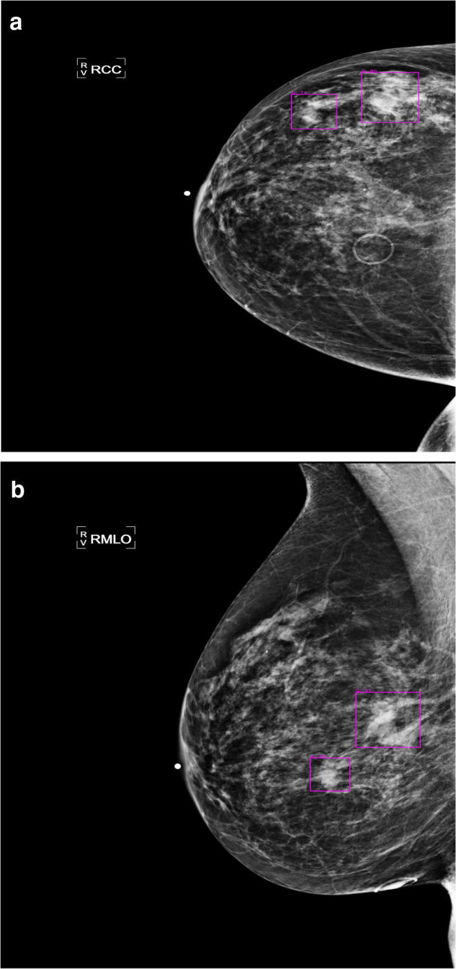
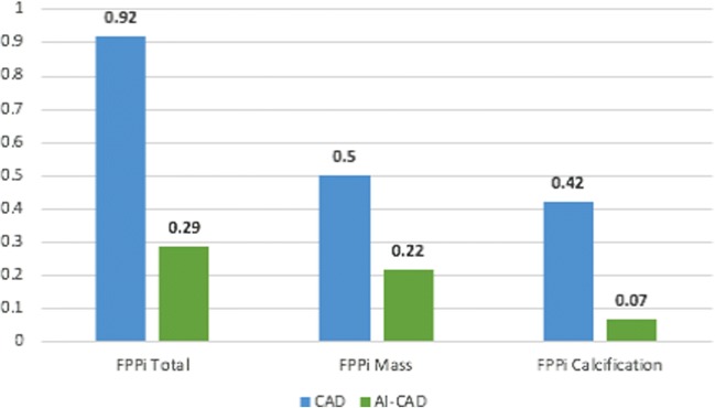
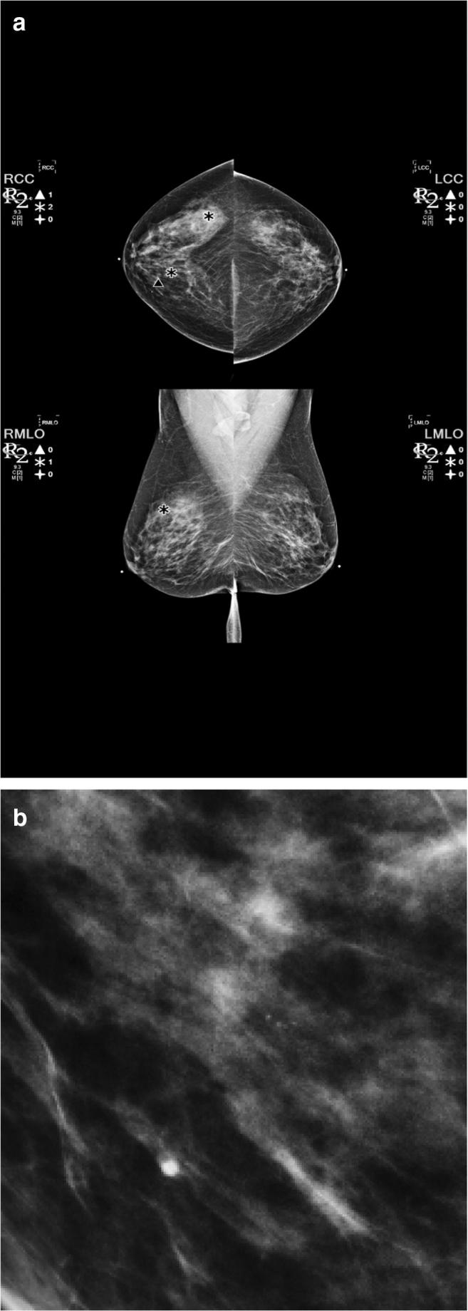
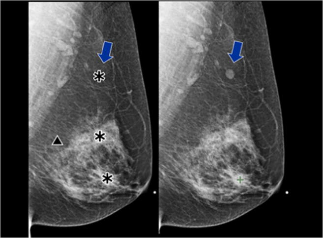
The aim was to determine whether an artificial intelligence (AI)-based, computer-aided detection (CAD) software can be used to reduce false positive per image (FPPI) on mammograms as compared to an FDA-approved conventional CAD. A retrospective study was performed on a set of 250 full-field digital mammograms between January 1, 2013, and March 31, 2013, and the number of marked regions of interest of two different systems was compared for sensitivity and specificity in cancer detection. The count of false-positive marks per image (FPPI) of the two systems was also evaluated as well as the number of cases that were completely mark-free. All results showed statistically significant reductions in false marks with the use of AI-CAD vs CAD (confidence interval = 95%) with no reduction in sensitivity. There is an overall 69% reduction in FPPI using the AI-based CAD as compared to CAD, consisting of 83% reduction in FPPI for calcifications and 56% reduction for masses. Almost half (48%) of cases showed no AI-CAD markings while only 17% show no conventional CAD marks. There was a significant reduction in FPPI with AI-CAD as compared to CAD for both masses and calcifications at all tissue densities. A 69% decrease in FPPI could result in a 17% decrease in radiologist reading time per case based on prior literature of CAD reading times. Additionally, decreasing false-positive recalls in screening mammography has many direct social and economic benefits.

摘要

目的在于确定人工智能(AI)辅助的计算机辅助检测(CAD)软件是否可用于减少全数字化乳腺摄影术(FFDM)中的每幅图像假阳性率(FPPI),与 FDA 批准的传统 CAD 相比。对 2013 年 1 月 1 日至 2013 年 3 月 31 日期间的一组 250 例全数字化乳腺摄影术进行了回顾性研究,比较了两种不同系统标记的感兴趣区域的数量,以评估其在癌症检测中的敏感性和特异性。还评估了两种系统的每幅图像假阳性标记数量(FPPI)以及完全无标记的病例数量。所有结果均表明,与 CAD 相比,使用 AI-CAD 可显著减少假阳性标记(置信区间为 95%),而不会降低敏感性。与 CAD 相比,使用基于 AI 的 CAD 可使 FPPI 总体减少 69%,其中钙化的 FPPI 减少 83%,肿块的 FPPI 减少 56%。几乎一半(48%)的病例没有 AI-CAD 标记,而只有 17%的病例没有传统 CAD 标记。在所有组织密度中,AI-CAD 与 CAD 相比,在肿块和钙化方面的 FPPI 均显著降低。基于 CAD 阅读时间的先前文献,FPPI 减少 69%可能导致每个病例的放射科医生阅读时间减少 17%。此外,减少筛查性乳腺摄影术的假阳性召回具有许多直接的社会效益和经济效益。

https://cdn.ncbi.nlm.nih.gov/pmc/blobs/e59d/6646646/5657edf48b60/10278_2018_168_Fig6_HTML.jpg
https://cdn.ncbi.nlm.nih.gov/pmc/blobs/e59d/6646646/62b8df5894d7/10278_2018_168_Fig1_HTML.jpg
https://cdn.ncbi.nlm.nih.gov/pmc/blobs/e59d/6646646/72bd0d625af0/10278_2018_168_Fig2_HTML.jpg
https://cdn.ncbi.nlm.nih.gov/pmc/blobs/e59d/6646646/2dfd8d643d85/10278_2018_168_Fig3_HTML.jpg
https://cdn.ncbi.nlm.nih.gov/pmc/blobs/e59d/6646646/3ba4f44b0fc9/10278_2018_168_Fig4_HTML.jpg
https://cdn.ncbi.nlm.nih.gov/pmc/blobs/e59d/6646646/efe3b198f99a/10278_2018_168_Fig5_HTML.jpg
https://cdn.ncbi.nlm.nih.gov/pmc/blobs/e59d/6646646/5657edf48b60/10278_2018_168_Fig6_HTML.jpg

相似文献

1
Reduction of False-Positive Markings on Mammograms: a Retrospective Comparison Study Using an Artificial Intelligence-Based CAD.基于人工智能 CAD 的回顾性对比研究:降低乳腺 X 线摄影中的假阳性标记
J Digit Imaging. 2019 Aug;32(4):618-624. doi: 10.1007/s10278-018-0168-6.
2
Improved Cancer Detection Using Artificial Intelligence: a Retrospective Evaluation of Missed Cancers on Mammography.利用人工智能提高癌症检测率:对乳腺 X 光摄影术漏诊癌症的回顾性评估。
J Digit Imaging. 2019 Aug;32(4):625-637. doi: 10.1007/s10278-019-00192-5.
3
A method to test the reproducibility and to improve performance of computer-aided detection schemes for digitized mammograms.一种用于测试数字化乳腺X线摄影计算机辅助检测方案的可重复性并提高其性能的方法。
Med Phys. 2004 Nov;31(11):2964-72. doi: 10.1118/1.1806291.
4
Computer-aided detection in digital mammography: false-positive marks and their reproducibility in negative mammograms.数字化乳腺摄影中的计算机辅助检测:阴性乳腺造影片中的假阳性标记及其可重复性
Acta Radiol. 2009 Nov;50(9):999-1004. doi: 10.3109/02841850903216700.
5
Screening mammography-detected cancers: sensitivity of a computer-aided detection system applied to full-field digital mammograms.乳腺钼靶筛查发现的癌症:应用于全视野数字化乳腺钼靶片的计算机辅助检测系统的敏感性
Radiology. 2007 Jul;244(1):104-11. doi: 10.1148/radiol.2441060756. Epub 2007 May 16.
6
Mammographic Surveillance After Breast-Conserving Therapy: Impact of Digital Breast Tomosynthesis and Artificial Intelligence-Based Computer-Aided Detection.保乳治疗后的乳腺 X 线摄影监测:数字乳腺断层合成和基于人工智能的计算机辅助检测的影响。
AJR Am J Roentgenol. 2022 Jan;218(1):42-51. doi: 10.2214/AJR.21.26506. Epub 2021 Aug 11.
7
Computer-aided detection system performance on current and previous digital mammograms in patients with contralateral metachronous breast cancer.计算机辅助检测系统对患对侧异时性乳腺癌患者当前及既往数字化乳腺钼靶影像的性能表现
Acta Radiol. 2012 May 1;53(4):376-81. doi: 10.1258/ar.2012.110521. Epub 2012 Mar 8.
8
Dual system approach to computer-aided detection of breast masses on mammograms.乳腺钼靶片上乳腺肿块计算机辅助检测的双系统方法。
Med Phys. 2006 Nov;33(11):4157-68. doi: 10.1118/1.2357838.
9
Effect of artificial intelligence-based computer-aided diagnosis on the screening outcomes of digital mammography: a matched cohort study.基于人工智能的计算机辅助诊断对数字乳腺 X 线摄影筛查结果的影响:一项匹配队列研究。
Eur Radiol. 2023 Oct;33(10):7186-7198. doi: 10.1007/s00330-023-09692-z. Epub 2023 May 16.
10
Comparison of two software versions of a commercially available computer-aided detection (CAD) system for detecting breast cancer.用于检测乳腺癌的一款商用计算机辅助检测(CAD)系统的两个软件版本的比较。
Acta Radiol. 2010 Jun;51(5):482-90. doi: 10.3109/02841851003709490.

引用本文的文献

1
AI-CAD-Guided Mammographic Assessment of Tumor Size and T Stage: Concordance with MRI for Clinical Staging in Breast Cancer Patients Considered for NAC.人工智能计算机辅助检测引导下对肿瘤大小和T分期的乳腺钼靶评估:与MRI在考虑接受新辅助化疗的乳腺癌患者临床分期中的一致性
Tomography. 2025 Jun 24;11(7):72. doi: 10.3390/tomography11070072.
2
Evaluating accountability, transparency, and bias in AI-assisted healthcare decision- making: a qualitative study of healthcare professionals' perspectives in the UK.评估人工智能辅助医疗决策中的问责制、透明度和偏差:对英国医疗专业人员观点的定性研究。
BMC Med Ethics. 2025 Jul 8;26(1):89. doi: 10.1186/s12910-025-01243-z.
3

本文引用的文献

1
Artificial intelligence and deep learning - Radiology's next frontier?人工智能与深度学习——放射学的下一个前沿领域?
Clin Imaging. 2018 May-Jun;49:87-88. doi: 10.1016/j.clinimag.2017.11.007. Epub 2017 Nov 16.
2
Screening for Breast Cancer: U.S. Preventive Services Task Force Recommendation Statement.乳腺癌筛查:美国预防服务工作组推荐声明。
Ann Intern Med. 2016 Feb 16;164(4):279-96. doi: 10.7326/M15-2886. Epub 2016 Jan 12.
3
Diagnostic Accuracy of Digital Screening Mammography With and Without Computer-Aided Detection.有和没有计算机辅助检测的数字化乳腺筛查钼靶摄影的诊断准确性
Curriculum check, 2025-equipping radiology residents for AI challenges of tomorrow.
课程检查,2025年——让放射科住院医师为未来的人工智能挑战做好准备。
Abdom Radiol (NY). 2025 Jun 9. doi: 10.1007/s00261-025-05016-5.
4
Diagnostic test accuracy of AI-assisted mammography for breast imaging: a narrative review.人工智能辅助乳腺钼靶摄影在乳腺成像中的诊断测试准确性:一项叙述性综述。
PeerJ Comput Sci. 2025 Feb 19;11:e2476. doi: 10.7717/peerj-cs.2476. eCollection 2025.
5
Traditional versus modern approaches to screening mammography: a comparison of computer-assisted detection for synthetic 2D mammography versus an artificial intelligence algorithm for digital breast tomosynthesis.传统与现代乳腺钼靶筛查方法:合成二维乳腺钼靶的计算机辅助检测与数字乳腺断层合成的人工智能算法之比较
Breast Cancer Res Treat. 2025 Apr;210(3):529-537. doi: 10.1007/s10549-024-07589-z. Epub 2025 Jan 9.
6
Assuring assistance to healthcare and medicine: Internet of Things, Artificial Intelligence, and Artificial Intelligence of Things.为医疗保健和医学提供保障:物联网、人工智能及物的人工智能。
Front Artif Intell. 2024 Dec 13;7:1442254. doi: 10.3389/frai.2024.1442254. eCollection 2024.
7
The Frontiers of Smart Healthcare Systems.智能医疗系统前沿
Healthcare (Basel). 2024 Nov 21;12(23):2330. doi: 10.3390/healthcare12232330.
8
Machine Learning Approaches for Skin Neoplasm Diagnosis.用于皮肤肿瘤诊断的机器学习方法
ACS Omega. 2024 Jul 15;9(30):32853-32863. doi: 10.1021/acsomega.4c03640. eCollection 2024 Jul 30.
9
Detection and Localization of Spine Disorders from Plain Radiography.通过X线平片检测和定位脊柱疾病
J Imaging Inform Med. 2024 Dec;37(6):2967-2982. doi: 10.1007/s10278-024-01175-x. Epub 2024 Jun 27.
10
Development of a Subtraction Processing Technology for Assistance in the Comparative Interpretation of Mammograms.一种用于辅助乳腺钼靶对比解读的减法处理技术的开发。
Diagnostics (Basel). 2024 May 29;14(11):1131. doi: 10.3390/diagnostics14111131.
JAMA Intern Med. 2015 Nov;175(11):1828-37. doi: 10.1001/jamainternmed.2015.5231.
4
The patient burden of screening mammography recall.筛查性乳房 X 光摄影召回的患者负担。
J Womens Health (Larchmt). 2014 Sep;23 Suppl 1:S11-9. doi: 10.1089/jwh.2014.1511.
5
Systematic review of the psychological consequences of false-positive screening mammograms.假阳性筛检乳房 X 光摄影的心理后果之系统性回顾。
Health Technol Assess. 2013 Mar;17(13):1-170, v-vi. doi: 10.3310/hta17130.
6
Computer-aided detection of masses at mammography: interactive decision support versus prompts.计算机辅助检测乳腺 X 线摄影中的肿块:交互式决策支持与提示。
Radiology. 2013 Jan;266(1):123-9. doi: 10.1148/radiol.12120218. Epub 2012 Oct 22.
7
Interpretation time of computer-aided detection at screening mammography.计算机辅助检测在乳腺 X 线筛查中的解读时间。
Radiology. 2010 Oct;257(1):40-6. doi: 10.1148/radiol.10092170. Epub 2010 Aug 2.
8
Influence of computer-aided detection on performance of screening mammography.计算机辅助检测对乳腺钼靶筛查性能的影响。
N Engl J Med. 2007 Apr 5;356(14):1399-409. doi: 10.1056/NEJMoa066099.
9
Prospective assessment of computer-aided detection in interpretation of screening mammography.在乳腺钼靶筛查解读中计算机辅助检测的前瞻性评估。
AJR Am J Roentgenol. 2006 Dec;187(6):1483-91. doi: 10.2214/AJR.05.1582.
10
Screening mammograms: interpretation with computer-aided detection--prospective evaluation.乳腺钼靶筛查:计算机辅助检测解读——前瞻性评估
Radiology. 2006 May;239(2):375-83. doi: 10.1148/radiol.2392042121. Epub 2006 Mar 28.