• 文献检索
  • 文档翻译
  • 深度研究
  • 学术资讯
  • Suppr Zotero 插件Zotero 插件
  • 邀请有礼
  • 套餐&价格
  • 历史记录
应用&插件
Suppr Zotero 插件Zotero 插件浏览器插件Mac 客户端Windows 客户端微信小程序
定价
高级版会员购买积分包购买API积分包
服务
文献检索文档翻译深度研究API 文档MCP 服务
关于我们
关于 Suppr公司介绍联系我们用户协议隐私条款
关注我们

Suppr 超能文献

核心技术专利:CN118964589B侵权必究
粤ICP备2023148730 号-1Suppr @ 2026

文献检索

告别复杂PubMed语法,用中文像聊天一样搜索,搜遍4000万医学文献。AI智能推荐,让科研检索更轻松。

立即免费搜索

文件翻译

保留排版,准确专业,支持PDF/Word/PPT等文件格式,支持 12+语言互译。

免费翻译文档

深度研究

AI帮你快速写综述,25分钟生成高质量综述,智能提取关键信息,辅助科研写作。

立即免费体验

传统与现代乳腺钼靶筛查方法:合成二维乳腺钼靶的计算机辅助检测与数字乳腺断层合成的人工智能算法之比较

Traditional versus modern approaches to screening mammography: a comparison of computer-assisted detection for synthetic 2D mammography versus an artificial intelligence algorithm for digital breast tomosynthesis.

作者信息

Bahl Manisha, Kshirsagar Ashwini, Pohlman Scott, Lehman Constance D

机构信息

Massachusetts General Hospital, 55 Fruit St, WAC 240, Boston, MA, 02114, USA.

Hologic, Inc., 250 Campus Drive, Marlborough, MA, 01752, USA.

出版信息

Breast Cancer Res Treat. 2025 Apr;210(3):529-537. doi: 10.1007/s10549-024-07589-z. Epub 2025 Jan 9.

DOI:10.1007/s10549-024-07589-z
PMID:39786500
原文链接:https://pmc.ncbi.nlm.nih.gov/articles/PMC11953105/
Abstract

PURPOSE

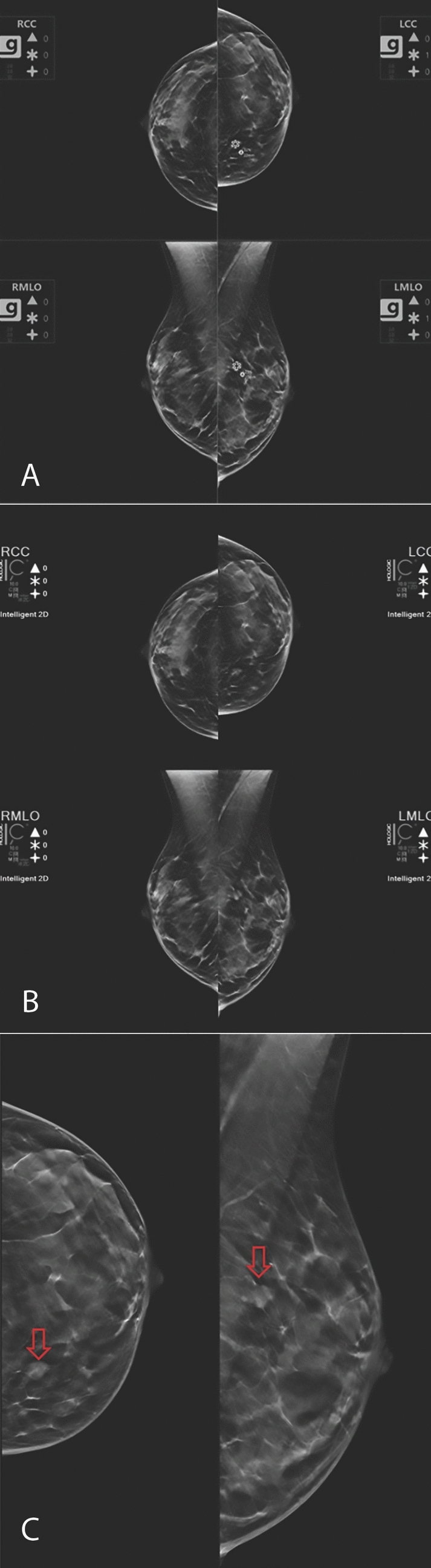
Traditional computer-assisted detection (CADe) algorithms were developed for 2D mammography, while modern artificial intelligence (AI) algorithms can be applied to 2D mammography and/or digital breast tomosynthesis (DBT). The objective is to compare the performance of a traditional machine learning CADe algorithm for synthetic 2D mammography to a deep learning-based AI algorithm for DBT on the same mammograms.

METHODS

Mammographic examinations from 764 patients (mean age 58 years ± 11) with 106 biopsy-proven cancers and 658 cancer-negative cases were analyzed by a CADe algorithm (ImageChecker v10.0, Hologic, Inc.) and an AI algorithm (Genius AI Detection v2.0, Hologic, Inc.). Synthetic 2D images were used for CADe analysis, and DBT images were used for AI analysis. For each algorithm, an overall case score was defined as the highest score of all lesion marks, which was used to determine the area under the receiver operating characteristic curve (AUC).

RESULTS

The overall AUC was higher for 3D AI than 2D CADe (0.873 versus 0.693, P < 0.001). Lesion-specific sensitivity of 3D AI was higher than 2D CADe (94.3 versus 72.6%, P = 0.002). Specificity of 3D AI was higher than 2D CADe (54.3 versus 16.7%, P < 0.001), and the rate of false marks on non-cancer cases was lower for 3D AI than 2D CADe (0.91 versus 3.24 per exam, P < 0.001).

CONCLUSION

A deep learning-based AI algorithm applied to DBT images significantly outperformed a traditional machine learning CADe algorithm applied to synthetic 2D mammographic images, with regard to AUC, sensitivity, and specificity.

摘要

目的

传统的计算机辅助检测(CADe)算法是为二维乳腺钼靶摄影开发的,而现代人工智能(AI)算法可应用于二维乳腺钼靶摄影和/或数字乳腺断层合成(DBT)。目的是在相同的乳房X光片上,比较用于合成二维乳腺钼靶摄影的传统机器学习CADe算法与用于DBT的基于深度学习的AI算法的性能。

方法

使用CADe算法(ImageChecker v10.0,Hologic公司)和AI算法(Genius AI Detection v2.0,Hologic公司)对764例患者(平均年龄58岁±11岁)的乳房X光检查进行分析,其中有106例经活检证实为癌症,658例为癌症阴性病例。合成二维图像用于CADe分析,DBT图像用于AI分析。对于每种算法,将总体病例评分定义为所有病变标记的最高分,用于确定受试者操作特征曲线(AUC)下的面积。

结果

三维AI的总体AUC高于二维CADe(0.873对0.693,P<0.001)。三维AI的病变特异性敏感性高于二维CADe(94.3%对72.6%,P=0.002)。三维AI的特异性高于二维CADe(54.3%对16.7%,P<0.001),三维AI在非癌症病例上的假标记率低于二维CADe(每次检查0.91对3.24,P<0.001)。

结论

在AUC、敏感性和特异性方面,应用于DBT图像的基于深度学习的AI算法明显优于应用于合成二维乳腺钼靶图像的传统机器学习CADe算法。

https://cdn.ncbi.nlm.nih.gov/pmc/blobs/45aa/11953105/f0fd4cc0a18c/10549_2024_7589_Fig3_HTML.jpg
https://cdn.ncbi.nlm.nih.gov/pmc/blobs/45aa/11953105/f563da17eee8/10549_2024_7589_Fig1_HTML.jpg
https://cdn.ncbi.nlm.nih.gov/pmc/blobs/45aa/11953105/f241dd6fbcfc/10549_2024_7589_Fig2_HTML.jpg
https://cdn.ncbi.nlm.nih.gov/pmc/blobs/45aa/11953105/f0fd4cc0a18c/10549_2024_7589_Fig3_HTML.jpg
https://cdn.ncbi.nlm.nih.gov/pmc/blobs/45aa/11953105/f563da17eee8/10549_2024_7589_Fig1_HTML.jpg
https://cdn.ncbi.nlm.nih.gov/pmc/blobs/45aa/11953105/f241dd6fbcfc/10549_2024_7589_Fig2_HTML.jpg
https://cdn.ncbi.nlm.nih.gov/pmc/blobs/45aa/11953105/f0fd4cc0a18c/10549_2024_7589_Fig3_HTML.jpg

相似文献

1
Traditional versus modern approaches to screening mammography: a comparison of computer-assisted detection for synthetic 2D mammography versus an artificial intelligence algorithm for digital breast tomosynthesis.传统与现代乳腺钼靶筛查方法:合成二维乳腺钼靶的计算机辅助检测与数字乳腺断层合成的人工智能算法之比较
Breast Cancer Res Treat. 2025 Apr;210(3):529-537. doi: 10.1007/s10549-024-07589-z. Epub 2025 Jan 9.
2
AI-enhanced Mammography With Digital Breast Tomosynthesis for Breast Cancer Detection: Clinical Value and Comparison With Human Performance.人工智能增强数字乳腺断层合成摄影术在乳腺癌检测中的应用:临床价值及与人类表现的比较。
Radiol Imaging Cancer. 2024 Jul;6(4):e230149. doi: 10.1148/rycan.230149.
3
Impact of AI for Digital Breast Tomosynthesis on Breast Cancer Detection and Interpretation Time.人工智能对数字乳腺断层合成术在乳腺癌检测和解读时间方面的影响。
Radiol Artif Intell. 2024 May;6(3):e230318. doi: 10.1148/ryai.230318.
4
Stand-Alone Use of Artificial Intelligence for Digital Mammography and Digital Breast Tomosynthesis Screening: A Retrospective Evaluation.人工智能在数字乳腺 X 线摄影和数字乳腺断层合成筛查中的独立应用:一项回顾性评估。
Radiology. 2022 Mar;302(3):535-542. doi: 10.1148/radiol.211590. Epub 2021 Dec 14.
5
External Testing of a Commercial AI Algorithm for Breast Cancer Detection at Screening Mammography.用于筛查乳腺钼靶摄影中乳腺癌检测的商用人工智能算法的外部测试
Radiol Artif Intell. 2025 May;7(3):e240287. doi: 10.1148/ryai.240287.
6
AI-based Strategies to Reduce Workload in Breast Cancer Screening with Mammography and Tomosynthesis: A Retrospective Evaluation.基于人工智能的策略可减少乳腺癌筛查中乳腺 X 线摄影和断层合成的工作量:回顾性评估。
Radiology. 2021 Jul;300(1):57-65. doi: 10.1148/radiol.2021203555. Epub 2021 May 4.
7
Impact of real-life use of artificial intelligence as support for human reading in a population-based breast cancer screening program with mammography and tomosynthesis.基于人群的乳腺癌筛查计划中,使用人工智能作为人类阅读支持的实际应用对乳腺 X 线摄影和断层合成的影响。
Eur Radiol. 2024 Jun;34(6):3958-3966. doi: 10.1007/s00330-023-10426-4. Epub 2023 Nov 17.
8
Mammographic Surveillance After Breast-Conserving Therapy: Impact of Digital Breast Tomosynthesis and Artificial Intelligence-Based Computer-Aided Detection.保乳治疗后的乳腺 X 线摄影监测:数字乳腺断层合成和基于人工智能的计算机辅助检测的影响。
AJR Am J Roentgenol. 2022 Jan;218(1):42-51. doi: 10.2214/AJR.21.26506. Epub 2021 Aug 11.
9
External Validation of a Commercial Artificial Intelligence Algorithm on a Diverse Population for Detection of False Negative Breast Cancers.一种用于检测假阴性乳腺癌的商业人工智能算法在不同人群中的外部验证
J Breast Imaging. 2025 Jan 25;7(1):16-26. doi: 10.1093/jbi/wbae058.
10
Performance of Digital Mammography-Based Artificial Intelligence Computer-Aided Diagnosis on Synthetic Mammography From Digital Breast Tomosynthesis.基于数字乳腺X线摄影的人工智能计算机辅助诊断在数字乳腺断层合成的合成乳腺X线摄影上的性能。
Korean J Radiol. 2025 Mar;26(3):217-229. doi: 10.3348/kjr.2024.0664.

引用本文的文献

1
Deep Learning with Transfer Learning on Digital Breast Tomosynthesis: A Radiomics-Based Model for Predicting Breast Cancer Risk.基于迁移学习的数字乳腺断层合成深度学习:一种基于影像组学的乳腺癌风险预测模型。
Diagnostics (Basel). 2025 Jun 26;15(13):1631. doi: 10.3390/diagnostics15131631.

本文引用的文献

1
Impact of a Categorical AI System for Digital Breast Tomosynthesis on Breast Cancer Interpretation by Both General Radiologists and Breast Imaging Specialists.分类人工智能系统对普通放射科医师和乳腺影像专家进行数字乳腺断层合成乳腺癌解读的影响。
Radiol Artif Intell. 2024 Mar;6(2):e230137. doi: 10.1148/ryai.230137.
2
Artificial Intelligence (AI) for Screening Mammography, From the Special Series on AI Applications.人工智能(AI)在乳腺 X 线摄影筛查中的应用,选自 AI 应用专题系列。
AJR Am J Roentgenol. 2022 Sep;219(3):369-380. doi: 10.2214/AJR.21.27071. Epub 2022 Jan 12.
3
Applications of Artificial Intelligence in Breast Imaging.
人工智能在乳腺成像中的应用。
Radiol Clin North Am. 2021 Jan;59(1):139-148. doi: 10.1016/j.rcl.2020.08.007.
4
Improving Accuracy and Efficiency with Concurrent Use of Artificial Intelligence for Digital Breast Tomosynthesis.同时使用人工智能提高数字乳腺断层合成的准确性和效率。
Radiol Artif Intell. 2019 Jul 31;1(4):e180096. doi: 10.1148/ryai.2019180096.
5
Reduction of False-Positive Markings on Mammograms: a Retrospective Comparison Study Using an Artificial Intelligence-Based CAD.基于人工智能 CAD 的回顾性对比研究:降低乳腺 X 线摄影中的假阳性标记
J Digit Imaging. 2019 Aug;32(4):618-624. doi: 10.1007/s10278-018-0168-6.
6
Breast Cancer Characteristics Associated with 2D Digital Mammography versus Digital Breast Tomosynthesis for Screening-detected and Interval Cancers.筛查检出癌和间期癌的 2D 数字乳腺钼钯与数字乳腺断层合成术的乳腺癌特征比较
Radiology. 2018 Apr;287(1):49-57. doi: 10.1148/radiol.2017171148. Epub 2017 Dec 22.
7
Clinical Performance of Synthesized Two-dimensional Mammography Combined with Tomosynthesis in a Large Screening Population.合成二维乳房 X 光摄影术与断层合成技术在大型筛查人群中的临床性能。
Radiology. 2017 Apr;283(1):70-76. doi: 10.1148/radiol.2017162674. Epub 2017 Feb 21.
8
Increased Cancer Detection Rate and Variations in the Recall Rate Resulting from Implementation of 3D Digital Breast Tomosynthesis into a Population-based Screening Program.将三维数字化乳腺断层合成技术应用于基于人群的筛查项目后,癌症检出率提高及召回率出现差异。
Radiology. 2016 Mar;278(3):698-706. doi: 10.1148/radiol.2015142036. Epub 2015 Oct 9.
9
Diagnostic Accuracy of Digital Screening Mammography With and Without Computer-Aided Detection.有和没有计算机辅助检测的数字化乳腺筛查钼靶摄影的诊断准确性
JAMA Intern Med. 2015 Nov;175(11):1828-37. doi: 10.1001/jamainternmed.2015.5231.
10
Breast cancer screening using tomosynthesis in combination with digital mammography.数字乳腺断层合成摄影术与数字乳腺钼靶摄影联合用于乳腺癌筛查。
JAMA. 2014 Jun 25;311(24):2499-507. doi: 10.1001/jama.2014.6095.