Institute of Pharmaceutical Sciences, ETH Zürich, Zurich, Switzerland.
Max Planck Institute for Molecular Biomedicine, Münster, Germany.
Front Immunol. 2019 Mar 22;10:520. doi: 10.3389/fimmu.2019.00520. eCollection 2019.
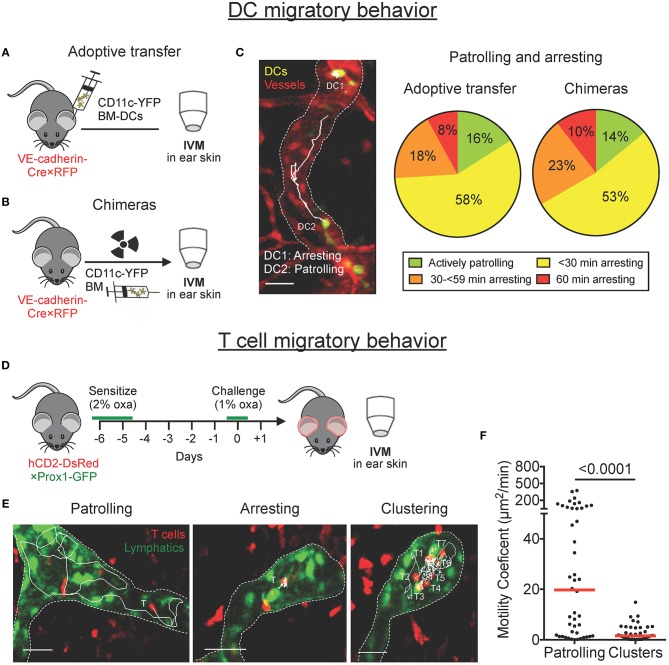
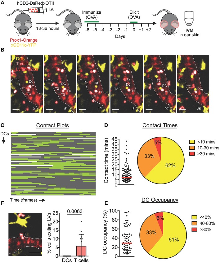
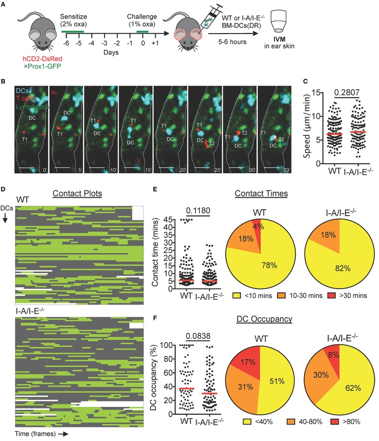
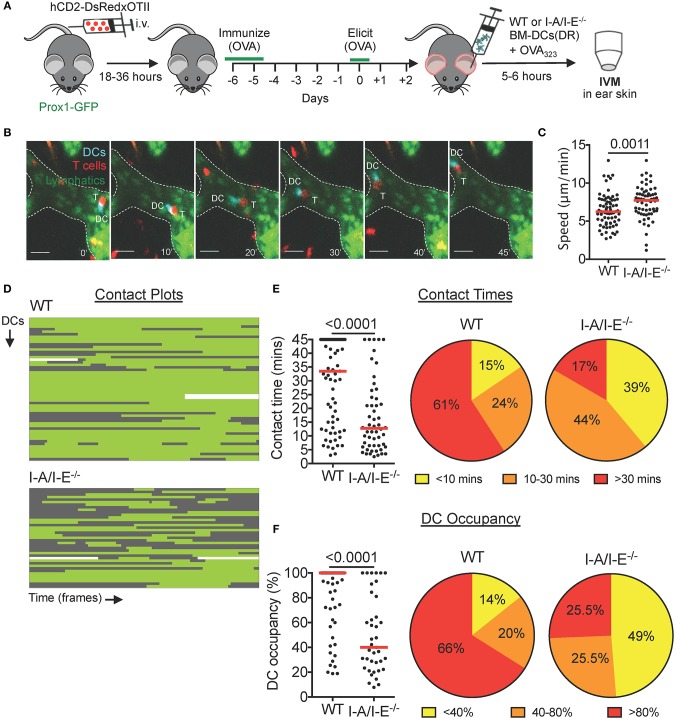
Afferent lymphatic vessels contribute to immunity by transporting antigen and leukocytes to draining lymph nodes (LNs) and are emerging as new players in the regulation of peripheral tolerance. Performing intravital microscopy in inflamed murine ear skin we found that migrating dendritic cells (DCs) and antigen-experienced effector T cells spend considerable time arresting or clustering within afferent lymphatic capillaries. We also observed that intralymphatic T cells frequently interacted with DCs. When imaging polyclonal T cells during an ongoing contact-hypersensitivity response, most intralymphatic DC-T cell interactions were short-lived. Conversely, during a delayed-type-hypersensitivity response, cognate antigen-bearing DCs engaged in long-lived MHCII-(I-A/I-E)-dependent interactions with antigen-specific T cells. Long-lived intralymphatic DC-T cell interactions reduced the speed of DC crawling but did not delay overall DC migration to draining LNs. While further consequences of these intralymphatic interactions still need to be explored, our findings suggest that lymphatic capillaries represent a unique compartment in which adaptive immune interaction and modulation occur.
Afferent lymphatic vessels contribute to immunity by transporting antigen and leukocytes to draining lymph nodes (LNs) and are emerging as new players in the regulation of peripheral tolerance. Performing intravital microscopy in inflamed murine ear skin we found that migrating dendritic cells (DCs) and antigen-experienced effector T cells spend considerable time arresting or clustering within afferent lymphatic capillaries. We also observed that intralymphatic T cells frequently interacted with DCs. When imaging polyclonal T cells during an ongoing contact-hypersensitivity response, most intralymphatic DC-T cell interactions were short-lived. Conversely, during a delayed-type-hypersensitivity response, cognate antigen-bearing DCs engaged in long-lived MHCII-(I-A/I-E)-dependent interactions with antigen-specific T cells. Long-lived intralymphatic DC-T cell interactions reduced the speed of DC crawling but did not delay overall DC migration to draining LNs. While further consequences of these intralymphatic interactions still need to be explored, our findings suggest that lymphatic capillaries represent a unique compartment in which adaptive immune interaction and modulation occur.
Afferent lymphatic vessels contribute to immunity by transporting antigen and leukocytes to draining lymph nodes (LNs) and are emerging as new players in the regulation of peripheral tolerance. Performing intravital microscopy in inflamed murine ear skin we found that migrating dendritic cells (DCs) and antigen-experienced effector T cells spend considerable time arresting or clustering within afferent lymphatic capillaries. We also observed that intralymphatic T cells frequently interacted with DCs. When imaging polyclonal T cells during an ongoing contact-hypersensitivity response, most intralymphatic DC-T cell interactions were short-lived. Conversely, during a delayed-type-hypersensitivity response, cognate antigen-bearing DCs engaged in long-lived MHCII-(I-A/I-E)-dependent interactions with antigen-specific T cells. Long-lived intralymphatic DC-T cell interactions reduced the speed of DC crawling but did not delay overall DC migration to draining LNs. While further consequences of these intralymphatic interactions still need to be explored, our findings suggest that lymphatic capillaries represent a unique compartment in which adaptive immune interaction and modulation occur.
传入的淋巴管通过将抗原和白细胞运输到引流淋巴结 (LNs) 来促进免疫,并且作为外周耐受调节的新参与者而出现。我们在发炎的鼠耳皮肤中进行活体显微镜检查,发现迁移的树突状细胞 (DC) 和抗原经验效应 T 细胞在传入淋巴管的毛细血管中花费相当多的时间停滞或聚集。我们还观察到淋巴管内的 T 细胞经常与 DC 相互作用。在进行中的接触超敏反应中对多克隆 T 细胞进行成像时,大多数淋巴管内 DC-T 细胞相互作用是短暂的。相反,在迟发型超敏反应中,携带同源抗原的 DC 与抗原特异性 T 细胞发生 MHCII-(I-A/I-E)-依赖性的长期相互作用。淋巴管内 DC-T 细胞的长期相互作用降低了 DC 爬行的速度,但并未延迟 DC 向引流 LNs 的整体迁移。虽然这些淋巴管内相互作用的进一步后果仍需要进一步探讨,但我们的研究结果表明,淋巴管代表一个独特的腔室,其中发生适应性免疫相互作用和调节。