Zhang Yufan, Zhang Juxiang, Li Xiaowei, Li Jingru, Lu Shuting, Li Yuqiao, Ren Panting, Zhang Chunfu, Xiong Liqin
Shanghai Med-X Engineering Center for Medical Equipment and Technology, School of Biomedical Engineering, Shanghai Jiao Tong University, Shanghai, 200030, PR China.
Mater Today Bio. 2022 Jun 12;15:100317. doi: 10.1016/j.mtbio.2022.100317. eCollection 2022 Jun.
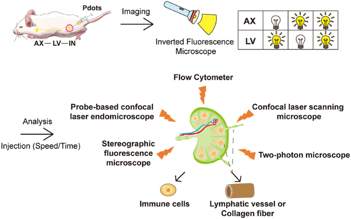
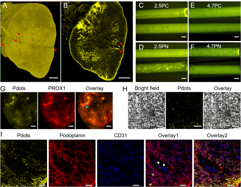
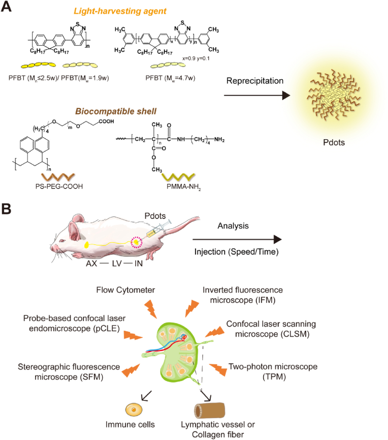
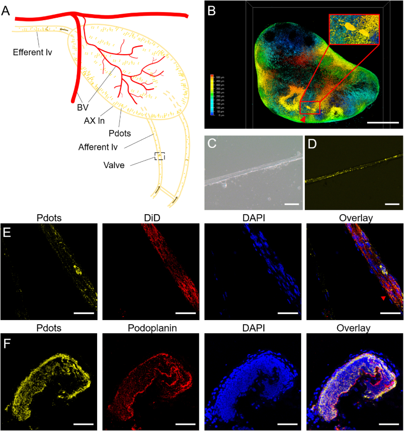
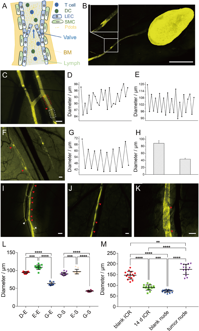
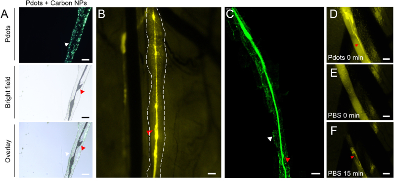
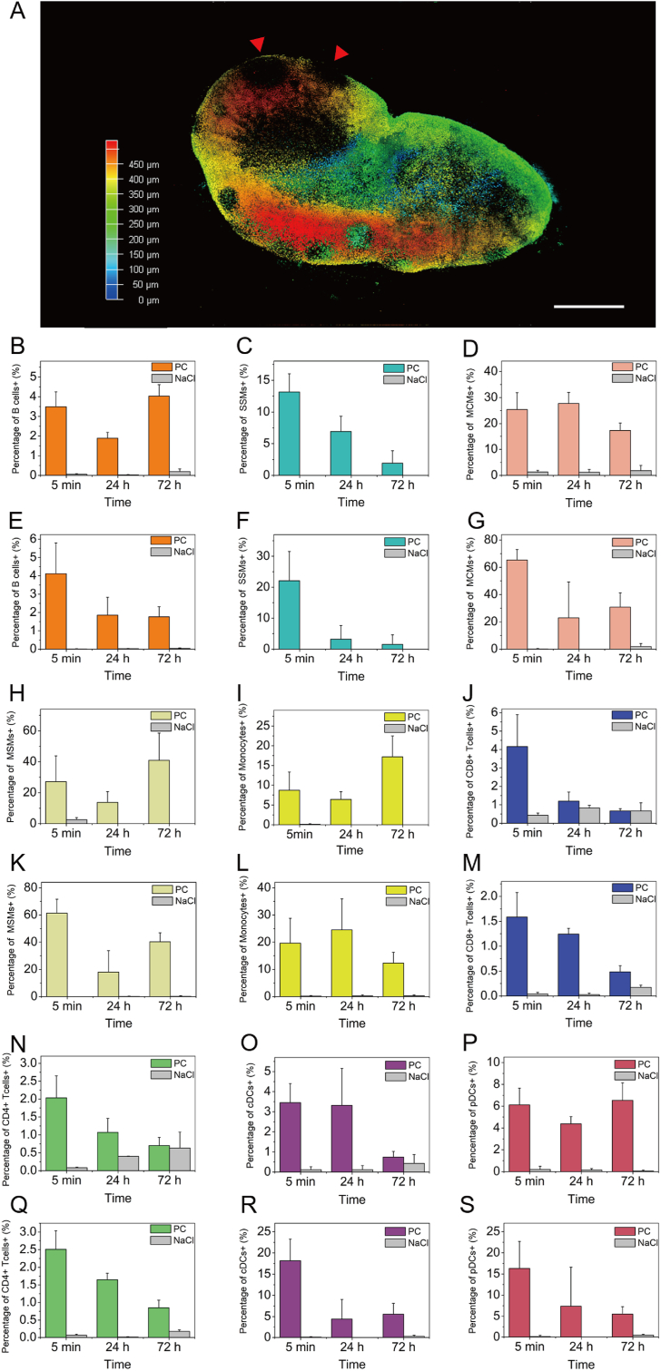
Polymer dots (Pdots) have been applied to imaging lymph nodes (LNs) and lymphatic vessels (LVs) in living mice and rats. However, the mechanism of absorption, distribution, metabolism, and excretion of Pdots in LNs and LVs is still unclear. Therefore, the relationship between Pdots and immune cells, LVs and collagen fibers in lymphatics was studied by multiple and microscopic imaging methods and detection techniques. Flow cytometry showed that Pdots could be phagocytosed by macrophages and monocytes, and had no relationship with B cells, T cells and dendric cells in LNs. Silver staining, immunofluorescence and two-photon microscope showed that Pdots gathered in collagen fibers and LVs of LNs. Furthermore, immunofluorescence imaging results verified that Pdots were distributed in the extracellular space of collecting LVs endothelial cells. In addition, Pdots in the collecting LVs were basically cleared by leaking into the surrounding tissue or draining LNs after 21 days of injection. During the long-time observation, Pdots also helped monitor the contraction frequency and variation range of LV. Our study lays a foundation on the research of Pdots as the carrier to study lymphatic structure and function in the future.
聚合物量子点(Pdots)已被应用于对活体小鼠和大鼠的淋巴结(LNs)及淋巴管(LVs)进行成像。然而,Pdots在LNs和LVs中的吸收、分布、代谢及排泄机制仍不清楚。因此,通过多种宏观和微观成像方法及检测技术,研究了Pdots与免疫细胞、LVs以及淋巴管中胶原纤维之间的关系。流式细胞术表明,Pdots可被巨噬细胞和单核细胞吞噬,与LNs中的B细胞、T细胞和树突状细胞无关。银染色、免疫荧光和双光子显微镜显示,Pdots聚集在LNs的胶原纤维和LVs中。此外,免疫荧光成像结果证实,Pdots分布在集合LVs内皮细胞的细胞外空间。另外,注射21天后,集合LVs中的Pdots基本通过渗漏到周围组织或引流到LNs而被清除。在长时间观察过程中,Pdots还有助于监测LV的收缩频率和变化范围。我们的研究为未来将Pdots作为研究淋巴结构和功能的载体奠定了基础。