Department of Radiology, Juntendo University Hospital, Tokyo, Japan.
Department of Radiology, Graduate School of Medicine, University of Tokyo, Tokyo, Japan.
J Magn Reson Imaging. 2019 Dec;50(6):1834-1842. doi: 10.1002/jmri.26744. Epub 2019 Apr 10.
Previous quantitative synthetic MRI of the brain has been solely performed in 2D.
To evaluate the feasibility of the recently developed sequence 3D-QALAS for brain cortical thickness and volumetric analysis.
Reproducibility/repeatability study.
Twenty-one healthy volunteers (35.6 ± 13.8 years).
FIELD STRENGTH/SEQUENCE: 3D T -weighted fast spoiled gradient recalled echo (FSPGR) sequence was performed once, and 3D-QALAS sequence was performed twice with a 3T scanner.
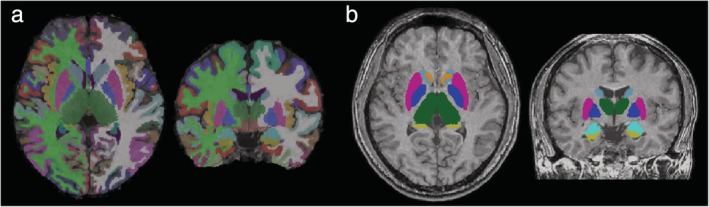
FreeSurfer and FIRST were used to measure cortical thickness and volume of subcortical structures, respectively. Agreement with FSPGR and scan-rescan repeatability were evaluated for 3D-QALAS.
Percent relative difference and intraclass correlation coefficient (ICC) were used to assess reproducibility and scan-rescan repeatability of the 3D-QALAS sequence-derived measurements.
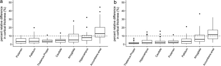
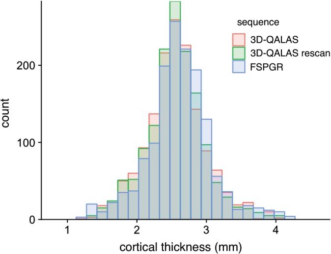
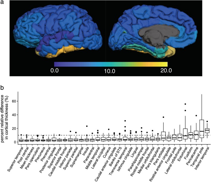
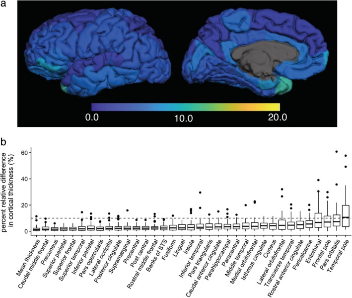
Percent relative difference compared with FSPGR in cortical thickness of the whole cortex was 3.1%, and 89% of the regional areas showed less than 10% relative difference in cortical thickness. The mean ICC across all regions was 0.65, and 74% of the structures showed substantial to almost perfect agreement. For volumes of subcortical structures, the median percent relative differences were lower than 10% across all subcortical structures, except for the accumbens area, and all structures showed ICCs of substantial to almost perfect agreement. For the scan-rescan test, percent relative difference in cortical thickness of the whole cortex was 2.3%, and 97% of the regional areas showed less than 10% relative difference in cortical thickness. The mean ICC across all regions was 0.73, and 80% showed substantial to almost perfect agreement. For volumes of subcortical structures, relative differences were less than 10% across all subcortical structures except for the accumbens area, and all structures showed ICCs of substantial to almost perfect agreement.
3D-QALAS could be reliably used for measuring cortical thickness and subcortical volumes in most brain regions.
3 Technical Efficacy: Stage 1 J. Magn. Reson. Imaging 2019;50:1834-1842.
之前的脑定量磁共振成像仅在 2D 下进行。
评估最近开发的序列 3D-QALAS 用于脑皮质厚度和容积分析的可行性。
再现/可重复性研究。
21 名健康志愿者(35.6±13.8 岁)。
磁场强度/序列:使用 3T 扫描仪进行一次 3D T1 加权快速扰相梯度回波(FSPGR)序列,两次 3D-QALAS 序列。
分别使用 FreeSurfer 和 FIRST 测量皮质下结构的皮质厚度和体积。评估 3D-QALAS 与 FSPGR 的一致性和扫描-重扫可重复性。
使用百分比相对差异和组内相关系数(ICC)评估 3D-QALAS 序列测量的再现性和扫描-重扫可重复性。
与 FSPGR 相比,皮质厚度的全脑皮质百分比相对差异为 3.1%,89%的区域皮质厚度相对差异小于 10%。所有区域的平均 ICC 为 0.65,74%的结构显示出较大到几乎完美的一致性。对于皮质下结构的体积,除了伏隔核区域外,所有皮质下结构的中位数百分比相对差异均低于 10%,所有结构的 ICC 均显示出较大到几乎完美的一致性。对于扫描-重扫测试,全脑皮质厚度的百分比相对差异为 2.3%,89%的区域皮质厚度相对差异小于 10%。所有区域的平均 ICC 为 0.73,80%显示出较大到几乎完美的一致性。对于皮质下结构的体积,除了伏隔核区域外,所有皮质下结构的相对差异均小于 10%,所有结构的 ICC 均显示出较大到几乎完美的一致性。
3D-QALAS 可用于可靠地测量大多数脑区的皮质厚度和皮质下体积。
3 技术功效:阶段 1 J. Magn. Reson. Imaging 2019;50:1834-1842.