Department of Radiology, Juntendo University, Tokyo, Japan.
Department of Radiology, The University of Tokyo, Tokyo, Japan.
Hum Brain Mapp. 2021 Feb 1;42(2):275-285. doi: 10.1002/hbm.25232. Epub 2020 Oct 22.
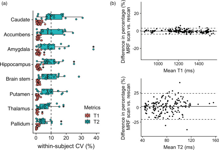
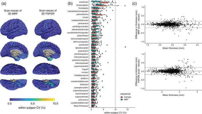
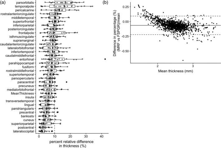
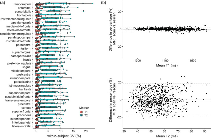
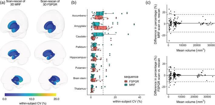
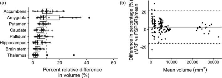
Three-dimensional (3D) Magnetic resonance fingerprinting (MRF) permits whole-brain volumetric quantification of T1 and T2 relaxation values, potentially replacing conventional T1-weighted structural imaging for common brain imaging analysis. The aim of this study was to evaluate the repeatability and reproducibility of 3D MRF in evaluating brain cortical thickness and subcortical volumetric analysis in healthy volunteers using conventional 3D T1-weighted images as a reference standard. Scan-rescan tests of both 3D MRF and conventional 3D fast spoiled gradient recalled echo (FSPGR) were performed. For each sequence, the regional cortical thickness and volume of the subcortical structures were measured using standard automatic brain segmentation software. Repeatability and reproducibility were assessed using the within-subject coefficient of variation (wCV), intraclass correlation coefficient (ICC), and mean percent difference and ICC, respectively. The wCV and ICC of cortical thickness were similar across all regions with both 3D MRF and FSPGR. The percent relative difference in cortical thickness between 3D MRF and FSPGR across all regions was 8.0 ± 3.2%. The wCV and ICC of the volume of subcortical structures across all structures were similar between 3D MRF and FSPGR. The percent relative difference in the volume of subcortical structures between 3D MRF and FSPGR across all structures was 7.1 ± 3.6%. 3D MRF measurements of human brain cortical thickness and subcortical volumes are highly repeatable, and consistent with measurements taken on conventional 3D T1-weighted images. A slight, consistent bias was evident between the two, and thus careful attention is required when combining data from MRF and conventional acquisitions.
三维(3D)磁共振指纹(MRF)可实现 T1 和 T2 弛豫值的全脑容积定量,可能替代常规 T1 加权结构成像用于常见的脑成像分析。本研究旨在评估 3D MRF 在使用常规 3D T1 加权图像作为参考标准评估健康志愿者脑皮质厚度和皮质下容积分析的重复性和可再现性。对 3D MRF 和常规 3D 快速扰相梯度回波(FSPGR)进行了扫描-再扫描测试。对于每个序列,使用标准的自动脑分割软件测量皮质区域的皮质厚度和皮质下结构的体积。使用个体内变异系数(wCV)、组内相关系数(ICC)和平均百分比差异和 ICC 分别评估重复性和可再现性。3D MRF 和 FSPGR 的所有区域的皮质厚度的 wCV 和 ICC 相似。3D MRF 和 FSPGR 之间所有区域的皮质厚度的相对百分比差异为 8.0±3.2%。3D MRF 和 FSPGR 的皮质下结构体积的 wCV 和 ICC 相似。3D MRF 和 FSPGR 之间所有结构的皮质下结构体积的相对百分比差异为 7.1±3.6%。3D MRF 测量的人脑皮质厚度和皮质下体积具有高度的可重复性,与常规 3D T1 加权图像上的测量结果一致。两种方法之间存在明显的、一致的偏差,因此在结合 MRF 和常规采集的数据时需要小心。