• 文献检索
  • 文档翻译
  • 深度研究
  • 学术资讯
  • Suppr Zotero 插件Zotero 插件
  • 邀请有礼
  • 套餐&价格
  • 历史记录
应用&插件
Suppr Zotero 插件Zotero 插件浏览器插件Mac 客户端Windows 客户端微信小程序
定价
高级版会员购买积分包购买API积分包
服务
文献检索文档翻译深度研究API 文档MCP 服务
关于我们
关于 Suppr公司介绍联系我们用户协议隐私条款
关注我们

Suppr 超能文献

核心技术专利:CN118964589B侵权必究
粤ICP备2023148730 号-1Suppr @ 2026

文献检索

告别复杂PubMed语法,用中文像聊天一样搜索,搜遍4000万医学文献。AI智能推荐,让科研检索更轻松。

立即免费搜索

文件翻译

保留排版,准确专业,支持PDF/Word/PPT等文件格式,支持 12+语言互译。

免费翻译文档

深度研究

AI帮你快速写综述,25分钟生成高质量综述,智能提取关键信息,辅助科研写作。

立即免费体验

基于多模态干扰的纳米结构和大分子运动成像揭示了紫外诱导的细胞突发。

Multimodal interference-based imaging of nanoscale structure and macromolecular motion uncovers UV induced cellular paroxysm.

机构信息

Department of Biomedical Engineering, Northwestern University, Evanston, IL, 60208, USA.

Department of Molecular Biosciences, Northwestern University, Evanston, IL, 60208, USA.

出版信息

Nat Commun. 2019 Apr 10;10(1):1652. doi: 10.1038/s41467-019-09717-6.

DOI:10.1038/s41467-019-09717-6
PMID:30971691
原文链接:https://pmc.ncbi.nlm.nih.gov/articles/PMC6458150/
Abstract

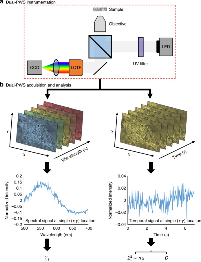
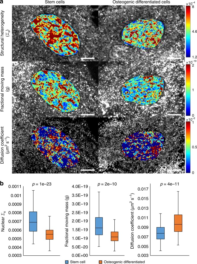
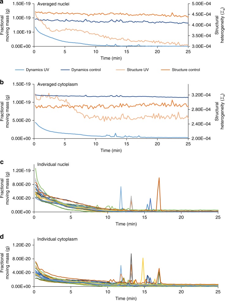
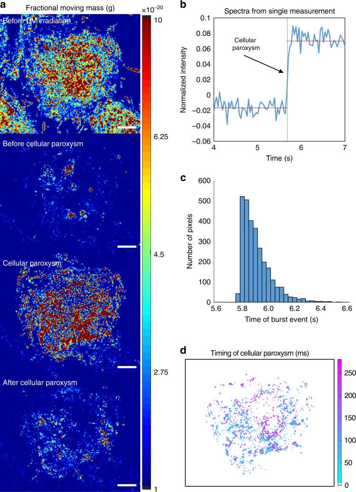
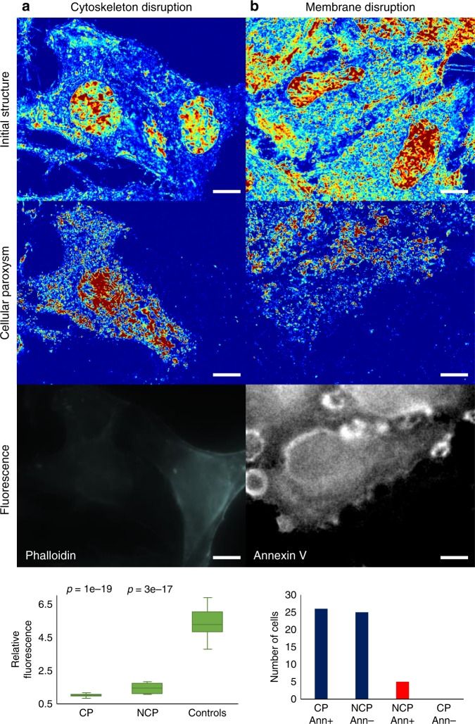
Understanding the relationship between intracellular motion and macromolecular structure remains a challenge in biology. Macromolecular structures are assembled from numerous molecules, some of which cannot be labeled. Most techniques to study motion require potentially cytotoxic dyes or transfection, which can alter cellular behavior and are susceptible to photobleaching. Here we present a multimodal label-free imaging platform for measuring intracellular structure and macromolecular dynamics in living cells with a sensitivity to macromolecular structure as small as 20 nm and millisecond temporal resolution. We develop and validate a theory for temporal measurements of light interference. In vitro, we study how higher-order chromatin structure and dynamics change during cell differentiation and ultraviolet (UV) light irradiation. Finally, we discover cellular paroxysms, a near-instantaneous burst of macromolecular motion that occurs during UV induced cell death. With nanoscale sensitive, millisecond resolved capabilities, this platform could address critical questions about macromolecular behavior in live cells.

摘要

理解细胞内运动与生物大分子结构之间的关系仍然是生物学领域的一项挑战。生物大分子结构是由众多分子组装而成的,其中一些分子无法被标记。大多数研究运动的技术都需要潜在细胞毒性的染料或转染,这可能会改变细胞行为,并且容易发生光漂白。在这里,我们提出了一种多模态无标记成像平台,用于测量活细胞内的结构和生物大分子动力学,对生物大分子结构的灵敏度低至 20nm,时间分辨率为毫秒级。我们开发并验证了一种用于光干涉时间测量的理论。在体外,我们研究了细胞分化和紫外线(UV)照射过程中高级染色质结构和动力学如何变化。最后,我们发现了细胞爆发,即在 UV 诱导的细胞死亡过程中发生的一种瞬间的生物大分子运动爆发。该平台具有纳米级的灵敏度和毫秒级的分辨率,能够解决关于活细胞中生物大分子行为的关键问题。

https://cdn.ncbi.nlm.nih.gov/pmc/blobs/369c/6458150/8758548f0df5/41467_2019_9717_Fig8_HTML.jpg
https://cdn.ncbi.nlm.nih.gov/pmc/blobs/369c/6458150/22472a380d28/41467_2019_9717_Fig1_HTML.jpg
https://cdn.ncbi.nlm.nih.gov/pmc/blobs/369c/6458150/e53dd2b35e54/41467_2019_9717_Fig2_HTML.jpg
https://cdn.ncbi.nlm.nih.gov/pmc/blobs/369c/6458150/65514b5b140f/41467_2019_9717_Fig3_HTML.jpg
https://cdn.ncbi.nlm.nih.gov/pmc/blobs/369c/6458150/835f6479bd66/41467_2019_9717_Fig4_HTML.jpg
https://cdn.ncbi.nlm.nih.gov/pmc/blobs/369c/6458150/3e7f1b084153/41467_2019_9717_Fig5_HTML.jpg
https://cdn.ncbi.nlm.nih.gov/pmc/blobs/369c/6458150/375325841c57/41467_2019_9717_Fig6_HTML.jpg
https://cdn.ncbi.nlm.nih.gov/pmc/blobs/369c/6458150/8ba53b9fe24a/41467_2019_9717_Fig7_HTML.jpg
https://cdn.ncbi.nlm.nih.gov/pmc/blobs/369c/6458150/8758548f0df5/41467_2019_9717_Fig8_HTML.jpg

相似文献

1
Multimodal interference-based imaging of nanoscale structure and macromolecular motion uncovers UV induced cellular paroxysm.基于多模态干扰的纳米结构和大分子运动成像揭示了紫外诱导的细胞突发。
Nat Commun. 2019 Apr 10;10(1):1652. doi: 10.1038/s41467-019-09717-6.
2
Label-free imaging of the native, living cellular nanoarchitecture using partial-wave spectroscopic microscopy.使用分波光谱显微镜对天然活细胞纳米结构进行无标记成像。
Proc Natl Acad Sci U S A. 2016 Oct 18;113(42):E6372-E6381. doi: 10.1073/pnas.1608198113. Epub 2016 Oct 4.
3
An orientation-independent DIC microscope allows high resolution imaging of epithelial cell migration and wound healing in a cnidarian model.一种与方向无关的 DIC 显微镜可用于在刺胞动物模型中对上皮细胞迁移和伤口愈合进行高分辨率成像。
J Microsc. 2018 Jun;270(3):290-301. doi: 10.1111/jmi.12682. Epub 2018 Jan 18.
4
A combined light sheet fluorescence and differential interference contrast microscope for live imaging of multicellular specimens.一种用于多细胞样本活体成像的联合光片荧光和微分干涉对比显微镜。
J Microsc. 2015 May;258(2):105-12. doi: 10.1111/jmi.12220. Epub 2015 Jan 22.
5
Scanning angle interference microscopy reveals cell dynamics at the nanoscale.扫描角干涉显微镜揭示纳米尺度的细胞动力学。
Nat Methods. 2012 Jul 1;9(8):825-7. doi: 10.1038/nmeth.2077.
6
Instantaneous Spatial Light Interference Microscopy.瞬时光学空间干涉显微镜
Opt Express. 2010 Jan 18;18(2):1569-75. doi: 10.1364/OE.18.001569.
7
Three-dimensional characterization of extreme ultraviolet mask blank defects by interference contrast photoemission electron microscopy.通过干涉对比光发射电子显微镜对极紫外掩膜版坯缺陷进行三维表征
Opt Express. 2008 Sep 29;16(20):15343-52. doi: 10.1364/oe.16.015343.
8
Photoregulated fluxional fluorophores for live-cell super-resolution microscopy with no apparent photobleaching.用于活细胞超分辨率显微镜的光调控荧光团,无明显光漂白。
Nat Commun. 2019 Mar 15;10(1):1232. doi: 10.1038/s41467-019-09217-7.
9
Nanoscale cellular imaging with scanning angle interference microscopy.利用扫描角干涉显微镜进行纳米级细胞成像。
Methods Cell Biol. 2014;123:235-52. doi: 10.1016/B978-0-12-420138-5.00013-6.
10
Structural dynamics of myosin 5 during processive motion revealed by interferometric scattering microscopy.干涉散射显微镜揭示的肌球蛋白5在持续运动过程中的结构动力学
Elife. 2015 Mar 6;4:e05413. doi: 10.7554/eLife.05413.

引用本文的文献

1
Nuclear blebs are associated with destabilized chromatin-packing domains.核泡与不稳定的染色质包装结构域相关。
J Cell Sci. 2025 Feb 1;138(3). doi: 10.1242/jcs.262161. Epub 2025 Feb 11.
2
Chromatin conformation, gene transcription, and nucleosome remodeling as an emergent system.染色质构象、基因转录和核小体重塑作为一个涌现系统。
Sci Adv. 2025 Jan 10;11(2):eadq6652. doi: 10.1126/sciadv.adq6652.
3
Decreased DNA density is a better indicator of a nuclear bleb than lamin B loss.与核纤层蛋白B缺失相比,DNA密度降低是核泡的更好指标。

本文引用的文献

1
Preservation of cellular nano-architecture by the process of chemical fixation for nanopathology.通过化学固定化纳米病理学的过程来保存细胞的纳米结构。
PLoS One. 2019 Jul 22;14(7):e0219006. doi: 10.1371/journal.pone.0219006. eCollection 2019.
2
Magnified Image Spatial Spectrum (MISS) microscopy for nanometer and millisecond scale label-free imaging.用于纳米和毫秒级无标记成像的放大图像空间光谱(MISS)显微镜技术。
Opt Express. 2018 Mar 5;26(5):5423-5440. doi: 10.1364/OE.26.005423.
3
Macrogenomic engineering via modulation of the scaling of chromatin packing density.
J Cell Sci. 2025 Feb 1;138(3). doi: 10.1242/jcs.262082. Epub 2025 Feb 11.
4
Hybrid Raman and Partial Wave Spectroscopy Microscope for the Characterization of Molecular and Structural Alterations in Tissue.用于表征组织中分子和结构变化的混合拉曼与分波光谱显微镜。
J Biophotonics. 2024 Dec;17(12):e202400330. doi: 10.1002/jbio.202400330. Epub 2024 Oct 27.
5
Probing chromatin condensation dynamics in live cells using interferometric scattering correlation spectroscopy.利用干涉散射相关光谱技术探测活细胞中的染色质凝聚动力学。
Commun Biol. 2024 Jun 24;7(1):763. doi: 10.1038/s42003-024-06457-2.
6
Depletion of lamins B1 and B2 promotes chromatin mobility and induces differential gene expression by a mesoscale-motion-dependent mechanism.层粘连蛋白 B1 和 B2 的耗竭通过介尺度运动依赖的机制促进染色质的流动性,并诱导差异基因表达。
Genome Biol. 2024 Mar 22;25(1):77. doi: 10.1186/s13059-024-03212-y.
7
DNA density is a better indicator of a nuclear bleb than lamin B loss.与核纤层蛋白B缺失相比,DNA密度是核泡的更好指标。
bioRxiv. 2024 Feb 7:2024.02.06.579152. doi: 10.1101/2024.02.06.579152.
8
Eukaryotic Cell Membranes: Structure, Composition, Research Methods and Computational Modelling.真核细胞的膜结构、组成、研究方法和计算建模。
Int J Mol Sci. 2023 Jul 7;24(13):11226. doi: 10.3390/ijms241311226.
9
Chromatin reprogramming and bone regeneration in vitro and in vivo via the microtopography-induced constriction of cell nuclei.通过细胞核微形貌诱导的收缩实现体外和体内的染色质重编程和骨再生。
Nat Biomed Eng. 2023 Nov;7(11):1514-1529. doi: 10.1038/s41551-023-01053-x. Epub 2023 Jun 12.
10
Origins of subdiffractional contrast in optical coherence tomography.光学相干断层扫描中次衍射对比度的起源
Biomed Opt Express. 2021 May 26;12(6):3630-3642. doi: 10.1364/BOE.416572. eCollection 2021 Jun 1.
通过调节染色质堆积密度的缩放进行宏基因组工程。
Nat Biomed Eng. 2017 Nov;1(11):902-913. doi: 10.1038/s41551-017-0153-2. Epub 2017 Nov 6.
4
The effects of chemical fixation on the cellular nanostructure.化学固定对细胞纳米结构的影响。
Exp Cell Res. 2017 Sep 15;358(2):253-259. doi: 10.1016/j.yexcr.2017.06.022. Epub 2017 Jun 30.
5
Review of interferometric spectroscopy of scattered light for the quantification of subdiffractional structure of biomaterials.用于定量生物材料亚衍射结构的漫散射光干涉光谱学综述。
J Biomed Opt. 2017 Mar 1;22(3):30901. doi: 10.1117/1.JBO.22.3.030901.
6
The Global Relationship between Chromatin Physical Topology, Fractal Structure, and Gene Expression.染色质物理拓扑结构、分形结构与基因表达的全球关系。
Sci Rep. 2017 Jan 24;7:41061. doi: 10.1038/srep41061.
7
Label-free imaging of the native, living cellular nanoarchitecture using partial-wave spectroscopic microscopy.使用分波光谱显微镜对天然活细胞纳米结构进行无标记成像。
Proc Natl Acad Sci U S A. 2016 Oct 18;113(42):E6372-E6381. doi: 10.1073/pnas.1608198113. Epub 2016 Oct 4.
8
Phase correlation imaging of unlabeled cell dynamics.无标记细胞动力学的相位相关成像。
Sci Rep. 2016 Sep 12;6:32702. doi: 10.1038/srep32702.
9
Higher Order Chromatin Modulator Cohesin SA1 Is an Early Biomarker for Colon Carcinogenesis: Race-Specific Implications.高阶染色质调节剂黏连蛋白SA1是结肠癌发生的早期生物标志物:种族特异性影响
Cancer Prev Res (Phila). 2016 Nov;9(11):844-854. doi: 10.1158/1940-6207.CAPR-16-0054. Epub 2016 Aug 22.
10
Nanoscale refractive index fluctuations detected via sparse spectral microscopy.通过稀疏光谱显微镜检测到的纳米级折射率波动。
Biomed Opt Express. 2016 Feb 19;7(3):883-93. doi: 10.1364/BOE.7.000883. eCollection 2016 Mar 1.