Division of Cancer and Stem Cells, Tumour and Vascular Biology Laboratories, Cancer Biology, School of Medicine, Queen's Medical Centre, University of Nottingham, Nottingham, UK.
National Heart & Lung Institute, Imperial College London, London, UK.
Microcirculation. 2019 Aug;26(6):e12549. doi: 10.1111/micc.12549. Epub 2019 Jun 19.
Arteriolargenesis can be induced by concomitant stimulation of nitric Oxide (NO)-Angiopoietin receptor (Tie)-Vascular Endothelial Growth Factor (VEGF) signaling in the rat mesentery angiogenesis assay. We hypothesized that the same combination of exogenously added growth factors would also have a positive impact on arteriolargenesis and, consequently, the recovery of blood flow in a model of unilateral hindlimb ischemia.
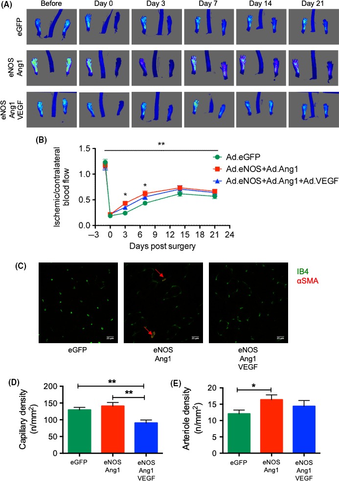
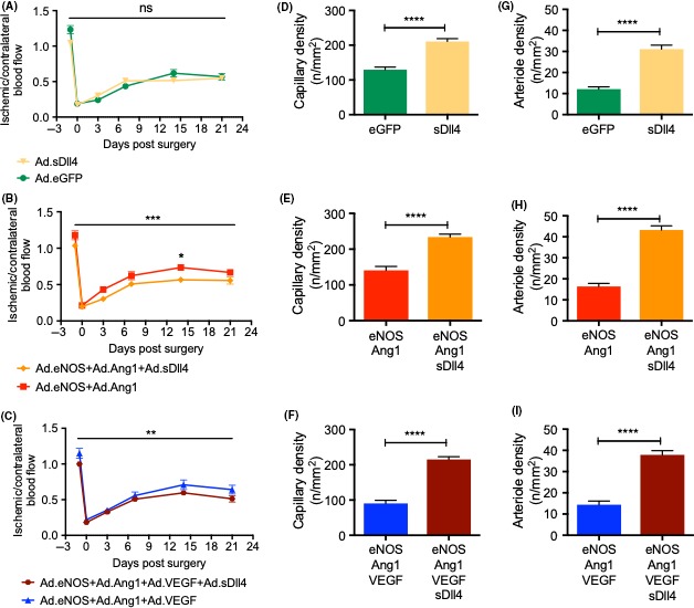
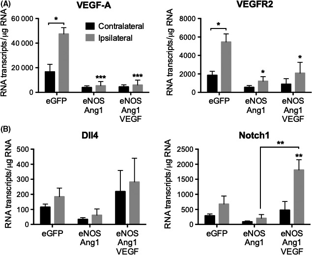
NO-Tie mice had faster blood flow recovery compared to control mice, as assessed by laser speckle imaging. There was no change in capillary density within the ischemic muscles, but arteriole density was higher in NO-Tie mice. Given the previously documented beneficial effect of VEGF signaling, we tested whether NO-Tie-VEGF mice would show further improvement. Surprisingly, these mice recovered no differently from control, arteriole density was similar and capillary density was lower. Dll4 is a driver of arterial specification, so we hypothesized that Notch1 expression would be involved in arteriolargenesis. There was a significant upregulation of Notch1 transcripts in NO-Tie-VEGF compared with NO-Tie mice. Using soluble Dll4 (sDll4), we stimulated Notch signaling in the ischemic muscles of mice. NO-Tie-sDll4 mice had significantly increased capillary and arteriole densities, but impaired blood flow recovery.
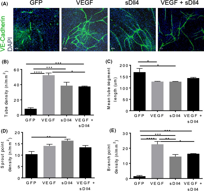
These results suggest that Dll4 activation early on in revascularization can lead to unproductive angiogenesis and arteriolargenesis, despite increased vascular densities. These results suggest spatial and temporal balance of growth factors needs to be perfected for ideal functional and anatomical revascularisation.
在大鼠肠系膜血管生成试验中,同时刺激一氧化氮(NO)-血管生成素受体(Tie)-血管内皮生长因子(VEGF)信号通路可诱导小动脉生成。我们假设,同样的外源性生长因子组合也会对小动脉生成产生积极影响,从而促进单侧后肢缺血模型中的血流恢复。
激光散斑成像评估表明,与对照组相比,NO-Tie 小鼠的血流恢复更快。缺血肌肉内的毛细血管密度没有变化,但 NO-Tie 小鼠的小动脉密度更高。鉴于之前报道的 VEGF 信号的有益作用,我们测试了 NO-Tie-VEGF 小鼠是否会进一步改善。令人惊讶的是,这些小鼠的恢复情况与对照组没有不同,小动脉密度相似,毛细血管密度较低。Dll4 是动脉特化的驱动因素,因此我们假设 Notch1 表达可能参与小动脉生成。与 NO-Tie 小鼠相比,NO-Tie-VEGF 小鼠中的 Notch1 转录物显著上调。使用可溶性 Dll4(sDll4),我们刺激了缺血肌肉中的 Notch 信号。NO-Tie-sDll4 小鼠的毛细血管和小动脉密度显著增加,但血流恢复受损。
这些结果表明,尽管血管密度增加,但血管生成和小动脉生成的早期 Dll4 激活可能导致无效的血管生成。这些结果表明,需要完善生长因子的时空平衡,以实现理想的功能和解剖血管重建。