The Wyss Institute for Biologically Inspired Engineering, Harvard University, Boston, Massachusetts, USA.
The Biological Design Center and Department of Biomedical Engineering, Boston University, Boston, Massachusetts, USA.
Nature. 2017 Dec 14;552(7684):258-262. doi: 10.1038/nature24998. Epub 2017 Nov 13.
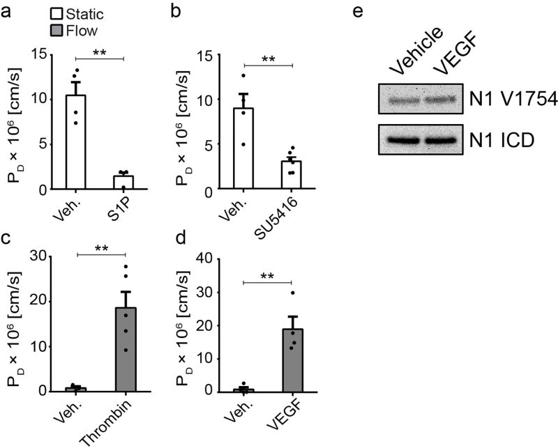
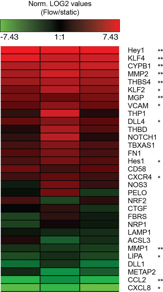
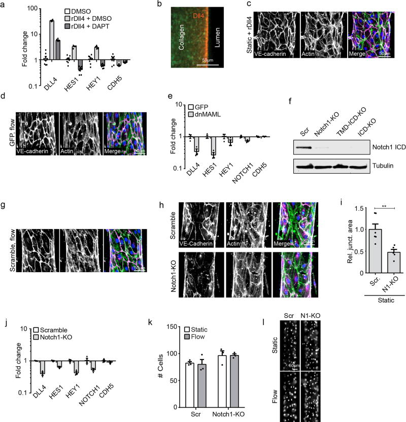
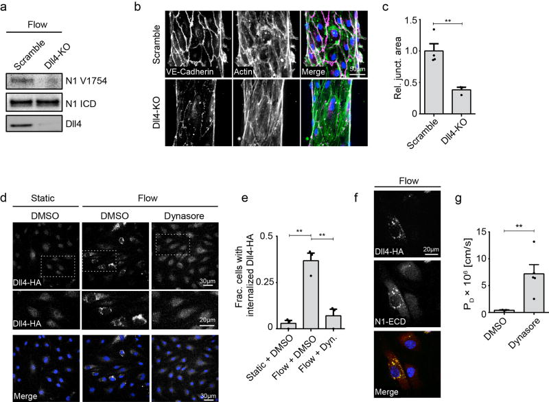
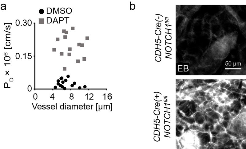
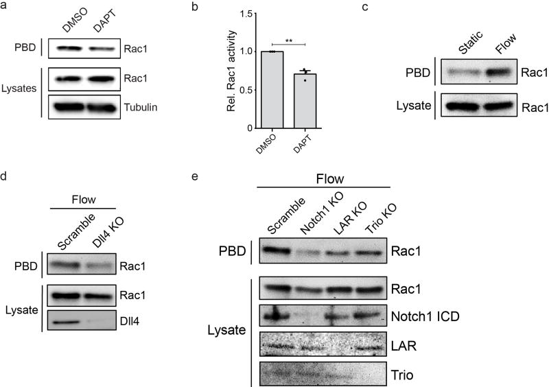
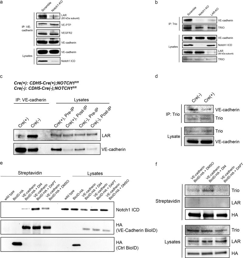
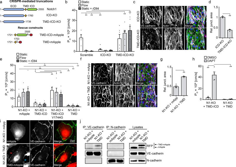
The vascular barrier that separates blood from tissues is actively regulated by the endothelium and is essential for transport, inflammation, and haemostasis. Haemodynamic shear stress plays a critical role in maintaining endothelial barrier function, but how this occurs remains unknown. Here we use an engineered organotypic model of perfused microvessels to show that activation of the transmembrane receptor NOTCH1 directly regulates vascular barrier function through a non-canonical, transcription-independent signalling mechanism that drives assembly of adherens junctions, and confirm these findings in mouse models. Shear stress triggers DLL4-dependent proteolytic activation of NOTCH1 to expose the transmembrane domain of NOTCH1. This domain mediates establishment of the endothelial barrier; expression of the transmembrane domain of NOTCH1 is sufficient to rescue defects in barrier function induced by knockout of NOTCH1. The transmembrane domain restores barrier function by catalysing the formation of a receptor complex in the plasma membrane consisting of vascular endothelial cadherin, the transmembrane protein tyrosine phosphatase LAR, and the RAC1 guanidine-exchange factor TRIO. This complex activates RAC1 to drive assembly of adherens junctions and establish barrier function. Canonical transcriptional signalling via Notch is highly conserved in metazoans and is required for many processes in vascular development, including arterial-venous differentiation, angiogenesis and remodelling. We establish the existence of a non-canonical cortical NOTCH1 signalling pathway that regulates vascular barrier function, and thus provide a mechanism by which a single receptor might link transcriptional programs with adhesive and cytoskeletal remodelling.
分隔血液和组织的血管屏障由内皮细胞主动调节,对于物质运输、炎症和止血至关重要。血流切应力在维持内皮屏障功能方面起着关键作用,但具体机制尚不清楚。本研究利用灌注微血管的工程化器官型模型,表明跨膜受体 NOTCH1 的激活通过一种非经典的、不依赖转录的信号机制直接调节血管屏障功能,该机制驱动黏附连接的组装,并在小鼠模型中证实了这些发现。切应力触发 DLL4 依赖性 NOTCH1 蛋白水解激活,从而暴露 NOTCH1 的跨膜结构域。该结构域介导内皮屏障的建立;NOTCH1 跨膜结构域的表达足以挽救 NOTCH1 敲除引起的屏障功能缺陷。跨膜结构域通过在质膜中催化包含血管内皮钙黏蛋白、跨膜蛋白酪氨酸磷酸酶 LAR 和 RAC1 鸟嘌呤交换因子 TRIO 的受体复合物的形成,恢复屏障功能。该复合物激活 RAC1 以驱动黏附连接的组装并建立屏障功能。经典的 Notch 转录信号在后生动物中高度保守,是血管发育过程中许多过程所必需的,包括动静脉分化、血管生成和重塑。本研究建立了一种非经典的皮质 NOTCH1 信号通路,该通路调节血管屏障功能,为单个受体将转录程序与黏附和细胞骨架重塑联系起来提供了一种机制。