Department of Ophthalmology and Visual Sciences, University of Michigan, Ann Arbor, MI, 48105, USA.
NTT-Hitech Insitute, Nguyen Tat Thanh University, Ho Chi Minh, Vietnam.
Sci Rep. 2019 Apr 11;9(1):5945. doi: 10.1038/s41598-019-42324-5.
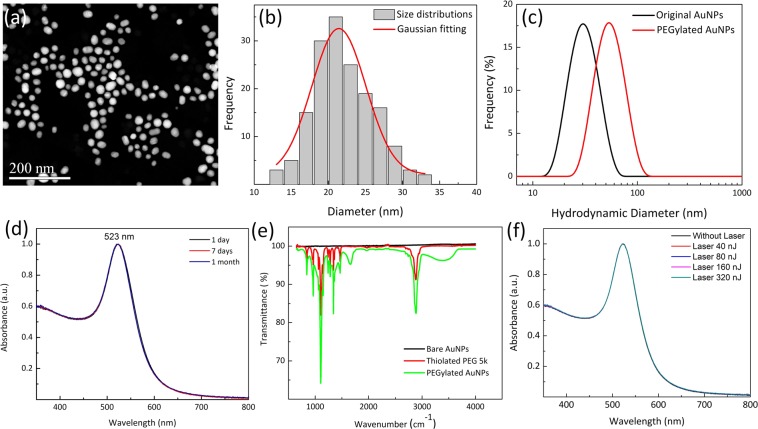
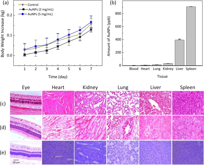
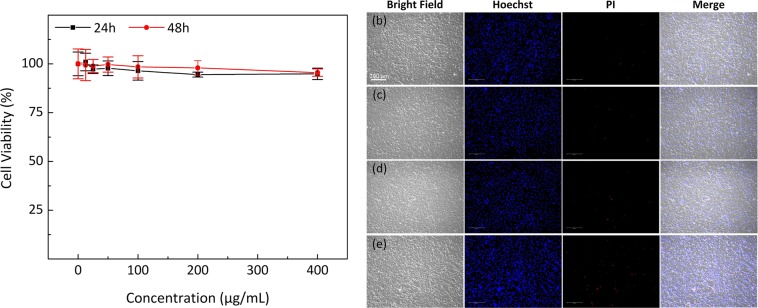
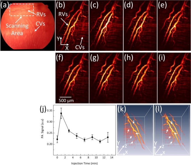
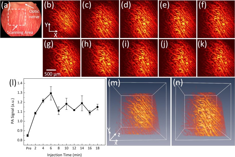
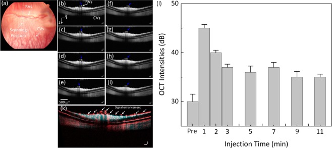
Multimodal imaging with photoacoustic microscopy (PAM) and optical coherence tomography (OCT) can be an effective method to evaluate the choroidal and retinal microvasculature. To improve the efficiency for visualizing capillaries, colloidal gold nanoparticles (AuNPs) have been applied as a multimodal contrast agent for both OCT and PAM imaging by taking advantage of the strong optical scattering and the strong optical absorption of AuNPs due to their surface plasmon resonance. Ultra-pure AuNPs were fabricated by femtosecond laser ablation, capped with polyethylene glycol (PEG), and administered to 13 New Zealand white rabbits and 3 Dutch Belted pigmented rabbits. The synthesized PEG-AuNPs (20.0 ± 1.5 nm) were demonstrated to be excellent contrast agents for PAM and OCT, and do not demonstrate cytotoxicity to bovine retinal endothelial cells in cell studies. The image signal from the retinal and choroidal vessels in living rabbits was enhanced by up to 82% for PAM and up to 45% for OCT, respectively, by the administered PEG-AuNPs, which enables detection of individual blood vessels by both imaging modalities. The biodistribution study demonstrated the AuNP accumulated primarily in the liver and spleen. Histology and TUNEL staining did not indicate cell injury or death in the lung, liver, kidney, spleen, heart, or eyes up to seven days after AuNP administration. PEG-AuNPs offer an efficient and safe contrast agent for multimodal ocular imaging to achieve improved characterization of microvasculature.
多模态成像技术结合光声显微镜(PAM)和光相干断层扫描(OCT)可以有效地评估脉络膜和视网膜的微血管。为了提高对毛细血管成像的效率,胶体金纳米颗粒(AuNPs)已被应用于 OCT 和 PAM 成像的多模态对比剂,这是利用 AuNPs 由于其表面等离子体共振而具有的强光学散射和强光学吸收特性。超纯 AuNPs 通过飞秒激光烧蚀制备,用聚乙二醇(PEG)进行包覆,并施用于 13 只新西兰白兔和 3 只荷兰带色兔。合成的 PEG-AuNPs(20.0±1.5nm)被证明是 PAM 和 OCT 的优异对比剂,在细胞研究中对牛视网膜内皮细胞没有显示出细胞毒性。在活体兔中,通过施用的 PEG-AuNPs,视网膜和脉络膜血管的 PAM 图像信号增强了 82%,OCT 图像信号增强了 45%,从而可以通过两种成像方式检测到单个血管。生物分布研究表明,AuNP 主要积聚在肝脏和脾脏中。组织学和 TUNEL 染色未表明在 AuNP 给药后 7 天内肺、肝、肾、脾、心或眼睛中存在细胞损伤或死亡。PEG-AuNPs 为多模态眼部成像提供了一种高效、安全的对比剂,以实现对微血管的更好特征化。