Ge Feifei, Mu Ping, Guo Rong, Cai Li, Liu Zheng, Dong Yan, Huang Yanhua H
Department of Psychiatry, University of Pittsburgh, Pittsburgh, PA, USA.
Department of Human Anatomy and Histoembryology, School of Medicine and Life Sciences, Nanjing University of Chinese Medicine, 210023, Nanjing, China.
Mol Psychiatry. 2021 Mar;26(3):941-954. doi: 10.1038/s41380-019-0419-z. Epub 2019 Apr 12.
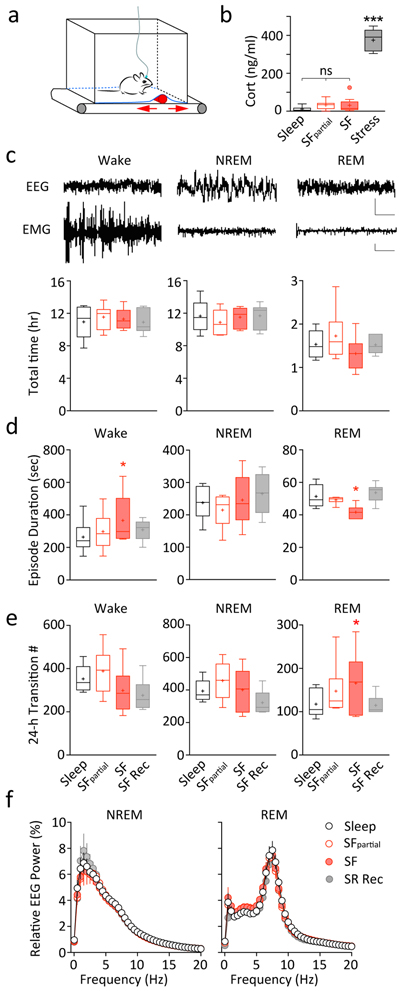
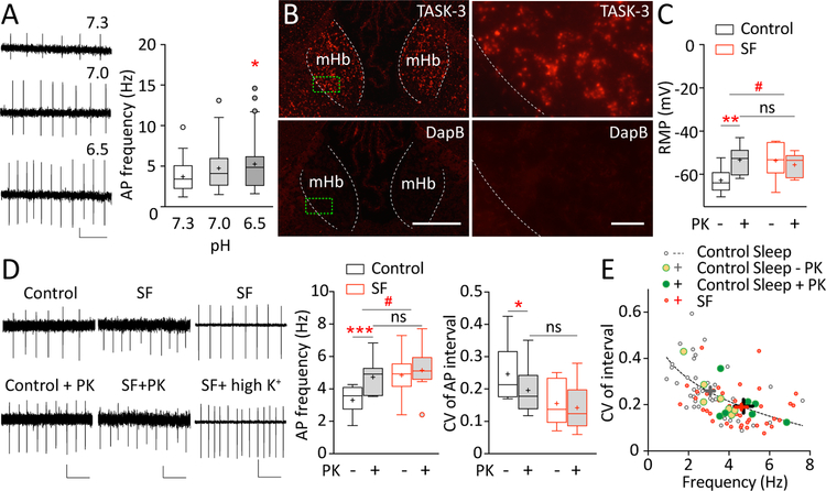
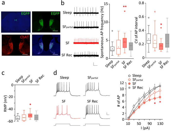
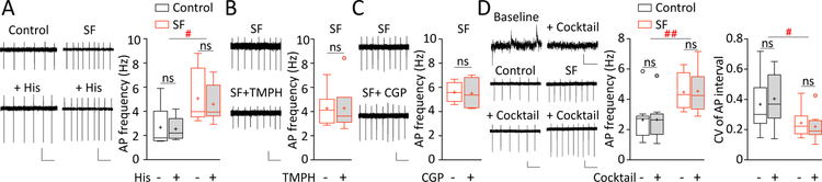
Sleep is essential to emotional health. Sleep disturbance, particularly REM sleep disturbance, profoundly impacts emotion regulation, but the underlying neural mechanisms remain elusive. Here we show that chronic REM sleep disturbance, achieved in mice by chronic sleep fragmentation (SF), enhanced neural activity in the medial habenula (mHb), a brain region increasingly implicated in negative affect. Specifically, after a 5-day SF procedure that selectively fragmented REM sleep, cholinergic output neurons (ChNs) in the mHb exhibited increased spontaneous firing rate and enhanced firing regularity in brain slices. The SF-induced firing changes remained intact upon inhibition of glutamate, GABA, acetylcholine, and histamine receptors, suggesting cell-autonomous mechanisms independent of synaptic transmissions. Moreover, the SF-induced hyperactivity was not because of enhanced intrinsic membrane excitability, but was accompanied by depolarized resting membrane potential in mHb ChNs. Furthermore, inhibition of TASK-3 (KCNK9) channels, a subtype of two-pore domain K channels, mimicked the SF effects by increasing the firing rate and regularity, as well as depolarizing the resting membrane potential in mHb ChNs in control-sleep mice. These effects of TASK-3 inhibition were absent in SF mice, suggesting reduced TASK-3 activity following SF. By contrast, inhibition of small-conductance Ca-activated K (SK) channels did not produce similar effects. Thus, SF compromised TASK-3 function in mHb ChNs, which likely led to depolarized resting membrane potential and increased spontaneous firing. These results not only demonstrate that selective REM sleep disturbance leads to hyperactivity of mHb ChNs, but also identify a key molecular substrate through which REM sleep disturbance may alter affect regulation.
睡眠对情绪健康至关重要。睡眠障碍,尤其是快速眼动(REM)睡眠障碍,会对情绪调节产生深远影响,但其潜在的神经机制仍不清楚。在这里,我们表明,通过慢性睡眠碎片化(SF)在小鼠中实现的慢性REM睡眠障碍,增强了内侧缰核(mHb)的神经活动,该脑区越来越多地与消极情绪有关。具体而言,在进行了5天选择性碎片化REM睡眠的SF程序后,mHb中的胆碱能输出神经元(ChNs)在脑片中表现出自发放电率增加和放电规律性增强。在抑制谷氨酸、γ-氨基丁酸、乙酰胆碱和组胺受体后,SF诱导的放电变化仍然存在,这表明细胞自主机制独立于突触传递。此外,SF诱导的多动并非由于内在膜兴奋性增强,而是伴随着mHb ChNs静息膜电位的去极化。此外,抑制双孔域钾通道亚型TASK-3(KCNK9)通道,通过增加放电率和规律性,以及使对照睡眠小鼠的mHb ChNs静息膜电位去极化,模拟了SF的作用。在SF小鼠中没有观察到TASK-3抑制的这些作用,这表明SF后TASK-3活性降低。相比之下,抑制小电导钙激活钾(SK)通道没有产生类似的效果。因此,SF损害了mHb ChNs中的TASK-3功能,这可能导致静息膜电位去极化和自发放电增加。这些结果不仅表明选择性REM睡眠障碍会导致mHb ChNs的多动,还确定了REM睡眠障碍可能改变情绪调节的关键分子底物。