• 文献检索
  • 文档翻译
  • 深度研究
  • 学术资讯
  • Suppr Zotero 插件Zotero 插件
  • 邀请有礼
  • 套餐&价格
  • 历史记录
应用&插件
Suppr Zotero 插件Zotero 插件浏览器插件Mac 客户端Windows 客户端微信小程序
定价
高级版会员购买积分包购买API积分包
服务
文献检索文档翻译深度研究API 文档MCP 服务
关于我们
关于 Suppr公司介绍联系我们用户协议隐私条款
关注我们

Suppr 超能文献

核心技术专利:CN118964589B侵权必究
粤ICP备2023148730 号-1Suppr @ 2026

文献检索

告别复杂PubMed语法,用中文像聊天一样搜索,搜遍4000万医学文献。AI智能推荐,让科研检索更轻松。

立即免费搜索

文件翻译

保留排版,准确专业,支持PDF/Word/PPT等文件格式,支持 12+语言互译。

免费翻译文档

深度研究

AI帮你快速写综述,25分钟生成高质量综述,智能提取关键信息,辅助科研写作。

立即免费体验

上颌窦的临床解剖学:在窦底增高术中的应用

Clinical anatomy of the maxillary sinus: application to sinus floor augmentation.

作者信息

Iwanaga Joe, Wilson Charlotte, Lachkar Stefan, Tomaszewski Krzysztof A, Walocha Jerzy A, Tubbs R Shane

机构信息

Seattle Science Foundation, Seattle, WA, USA.

Dental and Oral Medical Center, Kurume University School of Medicine, Kurume, Japan.

出版信息

Anat Cell Biol. 2019 Mar;52(1):17-24. doi: 10.5115/acb.2019.52.1.17. Epub 2019 Mar 29.

DOI:10.5115/acb.2019.52.1.17
PMID:30984447
原文链接:https://pmc.ncbi.nlm.nih.gov/articles/PMC6449588/
Abstract

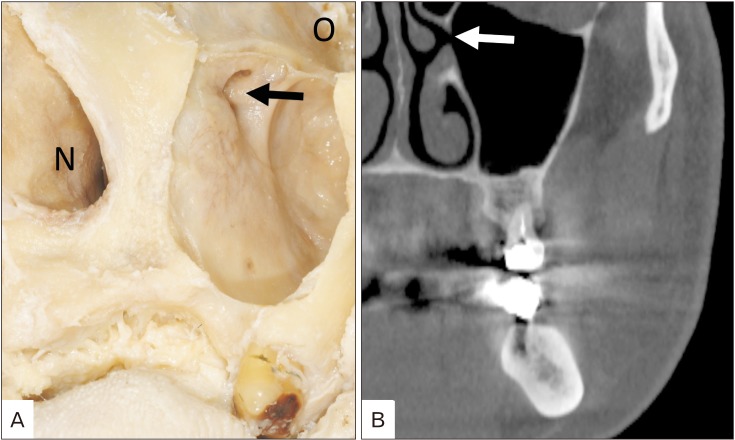
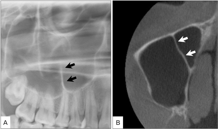
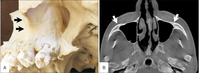
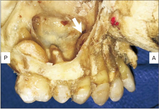
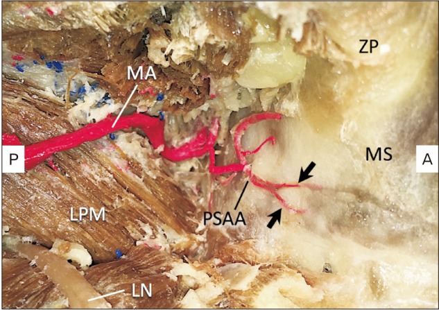
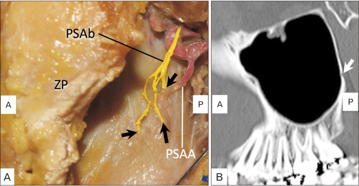
The anatomy of the maxillary sinus, especially its vascular anatomy, and its relationships with the teeth and alveolar processes have been well documented. The development of cone-beam computed tomography has resulted in dentists being more familiar with maxillary sinus floor augmentation procedures. This paper aims to revisit the classic anatomy of the maxillary sinus and review the newly published literature in order to help dentists diagnose in more detail and perform safer surgery of the maxillary sinus.

摘要

上颌窦的解剖结构,尤其是其血管解剖结构,以及它与牙齿和牙槽突的关系已有充分记载。锥形束计算机断层扫描技术的发展使牙医对上颌窦底提升手术更加熟悉。本文旨在重新审视上颌窦的经典解剖结构,并回顾新发表的文献,以帮助牙医更详细地进行诊断,并更安全地开展上颌窦手术。

https://cdn.ncbi.nlm.nih.gov/pmc/blobs/240b/6449588/27322f4c9480/acb-52-17-g006.jpg
https://cdn.ncbi.nlm.nih.gov/pmc/blobs/240b/6449588/86981a80c7f3/acb-52-17-g001.jpg
https://cdn.ncbi.nlm.nih.gov/pmc/blobs/240b/6449588/ff76a68eeaeb/acb-52-17-g002.jpg
https://cdn.ncbi.nlm.nih.gov/pmc/blobs/240b/6449588/b59377951d49/acb-52-17-g003.jpg
https://cdn.ncbi.nlm.nih.gov/pmc/blobs/240b/6449588/be61a3505416/acb-52-17-g004.jpg
https://cdn.ncbi.nlm.nih.gov/pmc/blobs/240b/6449588/d21ddc4255cd/acb-52-17-g005.jpg
https://cdn.ncbi.nlm.nih.gov/pmc/blobs/240b/6449588/27322f4c9480/acb-52-17-g006.jpg

相似文献

1
Clinical anatomy of the maxillary sinus: application to sinus floor augmentation.上颌窦的临床解剖学:在窦底增高术中的应用
Anat Cell Biol. 2019 Mar;52(1):17-24. doi: 10.5115/acb.2019.52.1.17. Epub 2019 Mar 29.
2
Sinus floor elevation or referral for further diagnosis and therapy: A comparison of maxillary sinus assessment by ENT specialists and dentists using cone beam computed tomography.上颌窦底提升或转专科进一步诊断和治疗:耳鼻喉科专家和牙医使用锥形束 CT 对上颌窦评估的比较。
Clin Oral Implants Res. 2020 May;31(5):463-475. doi: 10.1111/clr.13582. Epub 2020 Feb 12.
3
Radiological sinus lift: a new minimally invasive CT-guided procedure for maxillary sinus floor elevation in implant dentistry.放射学引导下的上颌窦提升术:种植牙领域中一种新型的微创CT引导下的上颌窦底提升手术。
Clin Oral Implants Res. 2016 Mar;27(3):341-7. doi: 10.1111/clr.12549. Epub 2015 Jan 13.
4
Anatomical analyses for maxillary sinus floor augmentation with a lateral approach: A cone beam computed tomography study.经外侧入路行上颌窦底提升的解剖学分析:锥形束 CT 研究。
Ann Anat. 2019 Nov;226:29-34. doi: 10.1016/j.aanat.2019.07.003. Epub 2019 Jul 19.
5
Long-term radiographic assessment of maxillary sinus floor augmentation using beta-tricalcium phosphate: analysis by cone-beam computed tomography.使用β-磷酸三钙对上颌窦底提升进行的长期影像学评估:锥形束计算机断层扫描分析
Int J Implant Dent. 2016 Dec;2(1):8. doi: 10.1186/s40729-016-0042-6. Epub 2016 Apr 1.
6
Sinus Floor Augmentation in Presence of Mucocele Eroding Maxillary Sinus Wall: A Case Report With 3 Years Follow-Up.上颌窦黏液囊肿侵蚀窦壁时的窦底提升:一例3年随访病例报告
Clin Adv Periodontics. 2020 Jun;10(2):81-87. doi: 10.1002/cap.10083. Epub 2019 Nov 15.
7
Evaluation of the Anastomosis Canal in Lateral Maxillary Sinus Wall With Cone Beam Computerized Tomography: A Clinical Study.锥形束计算机断层扫描评估上颌窦外侧壁吻合管:一项临床研究
J Oral Implantol. 2018 Feb;44(1):5-13. doi: 10.1563/aaid-joi-D-17-00129. Epub 2017 Oct 24.
8
Radiographic Evaluation of Maxillary Sinus Lateral Wall and Posterior Superior Alveolar Artery Anatomy: A Cone-Beam Computed Tomographic Study.上颌窦外侧壁及后上牙槽动脉解剖结构的影像学评估:一项锥形束计算机断层扫描研究
Clin Implant Dent Relat Res. 2017 Feb;19(1):151-160. doi: 10.1111/cid.12426. Epub 2016 May 30.
9
Evaluation of Single Tooth Loss to Maxillary Sinus and Surrounding Bone Anatomy With Cone-Beam Computed Tomography: A Multicenter Study.上颌窦及周围骨解剖中单牙缺失的锥形束 CT 评估:一项多中心研究。
Implant Dent. 2017 Oct;26(5):690-699. doi: 10.1097/ID.0000000000000652.
10
Optimized beagle model for maxillary sinus floor augmentation via a mini-lateral window with simultaneous implant placement.通过微型外侧窗口同时植入种植体进行上颌窦底提升的优化比格犬模型。
J Int Med Res. 2018 Nov;46(11):4684-4692. doi: 10.1177/0300060518796759. Epub 2018 Sep 10.

引用本文的文献

1
Relationship between zygomatic process morphology and maxillary sinus pneumatization.颧骨突形态与上颌窦气化之间的关系。
Saudi Dent J. 2025 Jul 1;37(4-6):25. doi: 10.1007/s44445-025-00031-0.
2
Using panoramic radiographs to assess the probability of causing oroantral communication following tooth removal. A retrospective cross-sectional study.使用全景X线片评估拔牙后发生口腔上颌窦瘘的可能性。一项回顾性横断面研究。
Clin Oral Investig. 2025 Apr 23;29(5):263. doi: 10.1007/s00784-025-06335-z.
3
Does the Anatomical Relationship Between Maxillary Molar Teeth and Maxillary Sinus Floor Differ Among Subjects with and Without Odontogenic Related Mucosal Thickening? A Cone-Beam Computed Tomographic Survey.

本文引用的文献

1
Three-dimensional CAD/CAM imaging of the maxillary sinus in ageing process.上颌窦在衰老过程中的三维CAD/CAM成像
Ann Anat. 2018 Jul;218:69-82. doi: 10.1016/j.aanat.2018.01.008. Epub 2018 Apr 5.
2
Comparison of cone-beam computed tomography and panoramic radiographs in detecting maxillary sinus septa.锥形束计算机断层扫描与全景X线片在上颌窦分隔检测中的比较。
J Istanb Univ Fac Dent. 2016 Oct 1;50(3):8-14. doi: 10.17096/jiufd.84476. eCollection 2016.
3
Prevalence and location of the posterior superior alveolar artery using cone-beam computed tomography.
在有无牙源性相关黏膜增厚的受试者中,上颌磨牙与上颌窦底之间的解剖关系是否存在差异?一项锥形束计算机断层扫描研究。
Indian J Otolaryngol Head Neck Surg. 2025 Apr;77(4):1782-1787. doi: 10.1007/s12070-025-05400-z. Epub 2025 Mar 12.
4
Discriminating between the maxillary tuberosity and the alveolar tuberosity- a critical pictorial review.区分上颌结节与牙槽结节——一项重要的影像学综述
Surg Radiol Anat. 2025 Jan 22;47(1):60. doi: 10.1007/s00276-025-03569-0.
5
Preoperative Cone Beam Computed Topography Assessment of Maxillary Sinus Variations in Dental Implant Patients.牙种植患者上颌窦变异的术前锥形束计算机断层扫描评估
Diagnostics (Basel). 2024 Sep 1;14(17):1929. doi: 10.3390/diagnostics14171929.
6
Anatomical relationship between maxillary posterior teeth and the maxillary sinus in an Indonesian population: a CT scan study.印度尼西亚人群上颌后牙与上颌窦的解剖关系:CT 扫描研究。
BMC Oral Health. 2024 Aug 29;24(1):1014. doi: 10.1186/s12903-024-04783-9.
7
Anatomy of Maxillary Sinus: Focus on Vascularization and Underwood Septa via 3D Imaging.上颌窦解剖结构:三维成像在血管化和安德伍德鼻中隔中的应用。
Tomography. 2024 Mar 24;10(4):444-458. doi: 10.3390/tomography10040034.
8
Automatic detection of posterior superior alveolar artery in dental cone-beam CT images using a deeply supervised multi-scale 3D network.利用深度监督多尺度 3D 网络自动检测口腔锥形束 CT 图像中的上颌后牙槽动脉。
Dentomaxillofac Radiol. 2024 Jan 11;53(1):22-31. doi: 10.1093/dmfr/twad002.
9
Resorbable magnesium metal membrane for sinus lift procedures: a case series.可吸收镁金属膜在鼻窦提升术中的应用:一组病例报告。
BMC Oral Health. 2023 Dec 14;23(1):1006. doi: 10.1186/s12903-023-03695-4.
10
Efficacy of mucolytics and steam therapy in the management of sinusitis among Indians.黏液溶解剂和蒸汽疗法对印度人鼻窦炎的治疗效果。
Bioinformation. 2023 Apr 30;19(4):479-483. doi: 10.6026/97320630019479. eCollection 2023.
使用锥形束计算机断层扫描技术观察后上牙槽动脉的患病率及位置
Imaging Sci Dent. 2017 Mar;47(1):39-44. doi: 10.5624/isd.2017.47.1.39. Epub 2017 Mar 21.
4
Evaluation of the position of the posterior superior alveolar artery in relation to the maxillary sinus using the Cone-Beam computed tomography scans.使用锥形束计算机断层扫描评估上颌后牙槽动脉相对于上颌窦的位置。
J Clin Exp Dent. 2017 Mar 1;9(3):e394-e399. doi: 10.4317/jced.53213. eCollection 2017 Mar.
5
Maxillary Sinus Dimensions Decrease as Age and Tooth Loss Increase.上颌窦尺寸随年龄增长和牙齿缺失增加而减小。
Implant Dent. 2017 Apr;26(2):288-295. doi: 10.1097/ID.0000000000000551.
6
Evaluation of the Posterior Superior Alveolar Artery Using Cone Beam Computed Tomography.使用锥形束计算机断层扫描评估上牙槽后动脉
Pol J Radiol. 2016 Dec 19;81:606-610. doi: 10.12659/PJR.899221. eCollection 2016.
7
Endoscopic management of the schneiderian membrane perforation during transcrestal sinus augmentation: a case report.经嵴状窦提升术中施耐德膜穿孔的内镜处理:一例报告
Oral Implantol (Rome). 2016 Nov 16;9(4):157-163. doi: 10.11138/orl/2016.9.4.157. eCollection 2016 Oct-Dec.
8
Transnasal Endoscopy Removal of Dislodged Dental Implant: A Case Report.经鼻内镜取出移位牙种植体:一例报告
J Oral Implantol. 2017 Jun;43(3):228-231. doi: 10.1563/aaid-joi-D-16-00172. Epub 2016 Dec 20.
9
Evaluation of the maxillary sinus in panoramic radiography-a comparative study.全景X线摄影中上颌窦的评估——一项对比研究。
Int J Implant Dent. 2015 Dec;1(1):17. doi: 10.1186/s40729-015-0015-1. Epub 2015 Jul 10.
10
Radiographic Evaluation of Maxillary Sinus Lateral Wall and Posterior Superior Alveolar Artery Anatomy: A Cone-Beam Computed Tomographic Study.上颌窦外侧壁及后上牙槽动脉解剖结构的影像学评估:一项锥形束计算机断层扫描研究
Clin Implant Dent Relat Res. 2017 Feb;19(1):151-160. doi: 10.1111/cid.12426. Epub 2016 May 30.