Private Practice, Tel Aviv, Israel.
Department of Dental Medicine, Faculty of Dental Medicine and Health Osijek, J.J. Strossmayer University of Osijek, Crkvena 21, 31 000, Osijek, Croatia.
BMC Oral Health. 2023 Dec 14;23(1):1006. doi: 10.1186/s12903-023-03695-4.
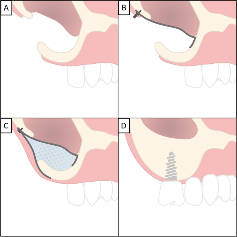
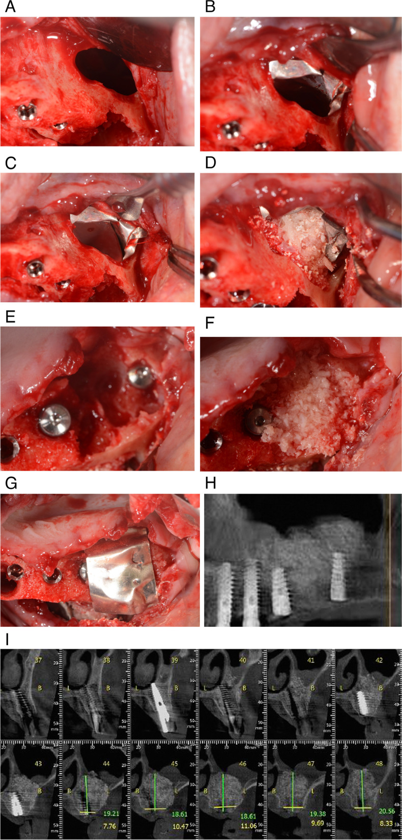
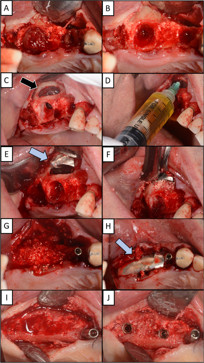
The purpose of this case series was to demonstrate the use of a magnesium membrane for repairing the perforated membrane in both direct and indirect approaches, as well as its application in instances where there has been a tear of the Schneiderian membrane.
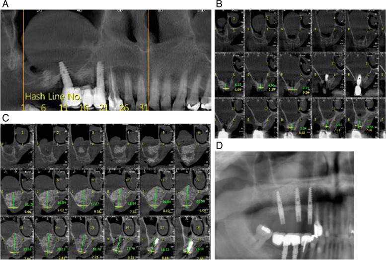
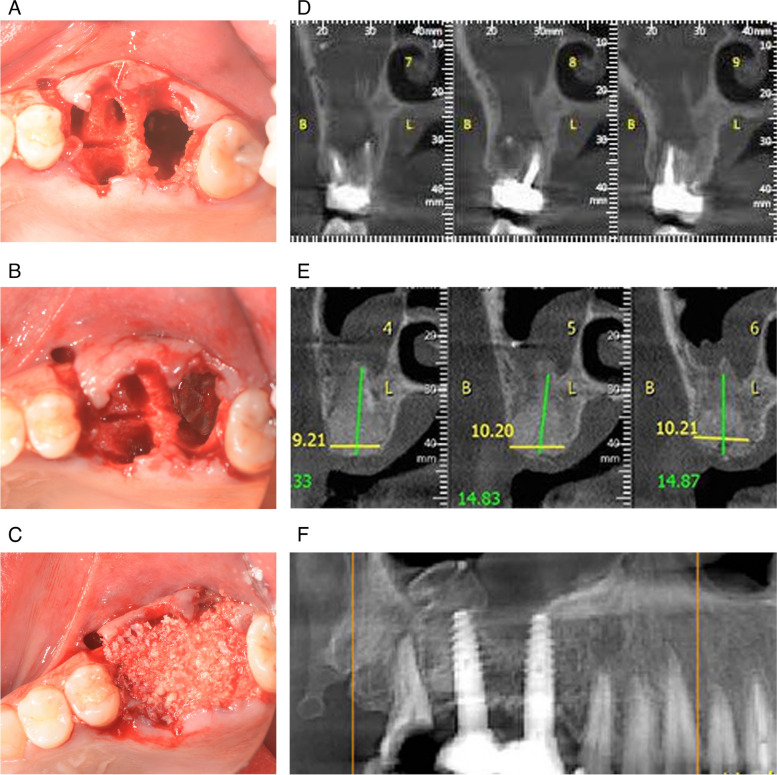
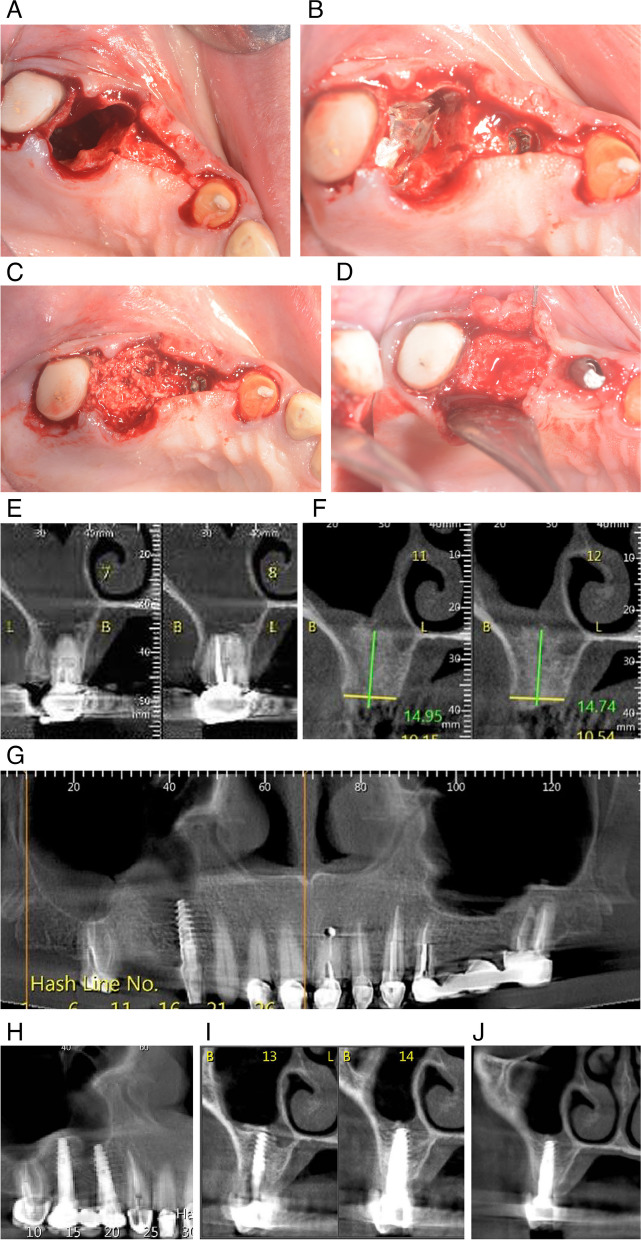
The case series included four individual cases, each demonstrating the application of a magnesium membrane followed by bone augmentation using a mixture of xenograft and allograft material in the sinus cavity. In the first three cases, rupture of Schneiderian membrane occurred as a result of tooth extraction, positioning of the dental implant, or as a complication during the procedure. In the fourth case, Schneiderian membrane was perforated as a result of the need to aspirate a polyp in the maxillary sinus. In case one, 10 mm of newly formed bone is visible four months after graft placement. Other cases showed between 15 and 20 mm of newly formed alveolar bone. No residual magnesium membrane was seen on clinical inspection. The vertical and horizontal augmentations proved stable and the dental implants were placed in the previously grafted sites.
Within the limitations of this case series, postoperative clinical examination, and panoramic and CBCT images demonstrated that resorbable magnesium membrane is a viable material for sinus lift and Schneiderian membrane repair. The case series showed successful healing and formation of new alveolar bone with separation of the oral cavity and maxillary sinus in four patients.
本病例系列旨在展示在直接和间接方法中使用镁膜修复穿孔膜的用途,以及在 Schneiderian 膜撕裂的情况下的应用。
该病例系列包括四个单独的病例,每个病例都展示了在鼻窦腔中使用异种移植物和同种移植物混合物进行骨增量后,再应用镁膜的情况。在前三个病例中,Schneiderian 膜破裂是由于拔牙、种植体定位或手术过程中的并发症引起的。在第四个病例中,Schneiderian 膜穿孔是因为需要抽吸上颌窦中的息肉。在第一个病例中,在放置移植物后四个月可看到 10 毫米的新骨形成。其他病例显示新牙槽骨形成了 15 到 20 毫米。临床检查未见残留的镁膜。垂直和水平增量证明是稳定的,并且种植体被放置在之前移植的部位。
在本病例系列的限制内,术后临床检查、全景和 CBCT 图像表明可吸收镁膜是鼻窦提升和Schneiderian 膜修复的一种可行材料。该病例系列显示了四名患者的口腔和上颌窦分离,成功愈合并形成了新的牙槽骨。