Shi Ce, Jiang Hong, Gameiro Giovana Rosa, Wang Jianhua
1School of Ophthalmology and Optometry, Wenzhou Medical University, Wenzhou, Zhejiang China.
2Bascom Palmer Eye Institute, University of Miami, Miller School of Medicine, 1638 NW 10th Avenue, McKnight Building - Room 202A, Miami, FL 33136 USA.
Eye Vis (Lond). 2019 Apr 6;6:11. doi: 10.1186/s40662-019-0136-3. eCollection 2019.
The aim was to determine the relationship between bulbar conjunctival microcirculation and retinal microcirculation in a healthy population.
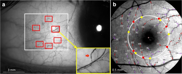
A functional slit-lamp biomicroscope (FSLB) was used to measure blood flow velocity (BFV) and blood flow rate (BFR) in the conjunctiva while a retinal function imager (RFI) was used to measure macular BFV and BFR in the retina. One eye of each subject of 58 self-reported healthy subjects was imaged in the same session on the same day.
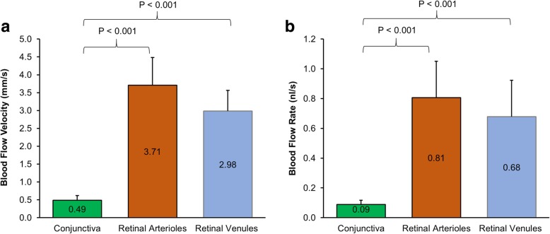
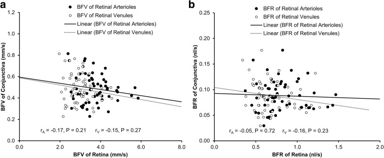
The mean BFV in the venules of the conjunctiva was 0.49 ± 0.13 mm/s, which was significantly slower than that in the retinal arterioles (3.71 ± 0.78 mm/s, < 0.001) and retinal venules (2.98 ± 0.58 mm/s, < 0.001). The BFR in the conjunctiva (0.09 nl/s) was also significantly lower than that in the retina (arterioles = 0.81 nl/s, venules = 0.68 nl/s, all < 0.001). The BFVs and BFRs were not related between the conjunctiva and retina (r ranged from - 0.17 to - 0.05, all > 0.05).
The microcirculation in the retina appeared to be different from that in the conjunctiva.
目的是确定健康人群中球结膜微循环与视网膜微循环之间的关系。
使用功能性裂隙灯生物显微镜(FSLB)测量结膜中的血流速度(BFV)和血流量(BFR),同时使用视网膜功能成像仪(RFI)测量视网膜中黄斑的BFV和BFR。在同一天的同一时段对58名自我报告健康的受试者的一只眼睛进行成像。
结膜小静脉中的平均BFV为0.49±0.13mm/s,明显慢于视网膜小动脉(3.71±0.78mm/s,<0.001)和视网膜小静脉(2.98±0.58mm/s,<0.001)中的BFV。结膜中的BFR(0.09nl/s)也明显低于视网膜中的BFR(小动脉=0.81nl/s,小静脉=0.68nl/s,均<0.001)。结膜和视网膜之间的BFV和BFR不相关(r范围为-0.17至-0.05,均>0.05)。
视网膜中的微循环似乎与结膜中的不同。