The Department of Human Genetics, Radboud University Medical Center, Nijmegen, the Netherlands (Hoogman, Guimaraes, Shumskaya, Wolfers, Bralten, Franke); the Donders Institute for Brain, Cognition, and Behavior, Radboud University, Nijmegen, the Netherlands (Hoogman, Shumskaya, Mennes, Wolfers, Buitelaar, Bralten, Franke); the Department of Child and Adolescent Psychiatry, Erasmus MC, University Medical Center Rotterdam, Rotterdam, the Netherlands (Muetzel, El Marroun, White, Tiemeier); the Department of Epidemiology, Erasmus MC, University Medical Center Rotterdam, Rotterdam, the Netherlands (Muetzel); the Department of Cognitive Neuroscience, Donders Institute for Brain, Cognition, and Behavior, Nijmegen, the Netherlands (Guimaraes, Zwiers, Buitelaar); the Imaging Genetics Center, Stevens Neuroimaging and Informatics Institute, Keck School of Medicine of USC, Marina del Rey, Calif. (Jahanshad, Thompson); National Human Genome Research Institute, Bethesda, Md. (Sudre, Shaw); the Department of Behavioral Neuroscience, Oregon Health and Science University, Portland (Earl, Fair, Nigg); the Department of Psychiatry and Forensic Medicine, Autonomous University of Barcelona, Cerdanyola del Vallès, Spain (Soliva Vila, Ramos-Quiroga, Vilarroya); Instituto ITACA, Polytechnic University of Valencia, Valencia, Spain (Vives-Gilabert); the Olin Neuropsychiatry Research Center, Hartford Hospital, Hartford, Conn. (Khadka, Novotny, Stevens); University of Groningen, University Medical Center Groningen (UMCG), Department of Psychiatry, Interdisciplinary Center Psychopathology and Emotion Regulation (ICPE), Groningen, the Netherlands (Hartman, Schweren); Faculty of Behavioral and Movement Sciences, Vrije Universiteit Amsterdam, Amsterdam (Heslenfeld); the Department of Child and Adolescent Psychiatry, University of Groningen, University Medical Center Groningen, the Netherlands (Hoekstra); NICHE Lab, Department of Psychiatry, UMC Utrecht Brain Center, Utrecht, the Netherlands (Ambrosino, Oranje, de Zeeuw, Durston); Laboratory of Psychiatric Neuroimaging (LIM-21), Department and Institute of Psychiatry, Faculty of Medicine, University of São Paulo, São Paulo, Brazil (Chaim-Avancini, Rosa, Zanetti, Busatto); the Center for Interdisciplinary Research on Applied Neurosciences (NAPNA), University of São Paulo, São Paulo, Brazil (Chaim-Avancini, Rosa, Zanetti, Busatto); the Developmental Imaging Group, Murdoch Children's Research Institute, Melbourne, Australia (Malpas); the Clinical Outcomes Research Unit (CORe), Department of Medicine, Royal Melbourne Hospital, University of Melbourne, Melbourne, Australia (Malpas); the Melbourne School of Psychological Sciences, University of Melbourne, Melbourne, Australia (Malpas); the Child Neuropsychology Section, University Hospital RWTH Aachen, Aachen, Germany (Kohls, Konrad; Child and Adolescent Psychiatry, University Hospital RWTH Aachen, Aachen, Germany (Polier, Seitz); Institute of Neuroscience and Medicine-Brain and Behavior (INM-7), Research Center Jülich, Jülich, Germany (Polier); the Clinical and Research Programs in Pediatric Psychopharmacology and Adult ADHD, Department of Psychiatry, Massachusetts General Hospital, Boston (Biederman); the Department of Psychiatry, Massachusetts General Hospital, Harvard Medical School, Boston (Biederman, Doyle); the Center for Genomic Medicine, Massachusetts General Hospital, Harvard Medical School, Boston (Doyle); the Departments of Neurosciences, Radiology, and Psychiatry and the Center for Multimodal Imaging and Genetics, University of California San Diego (Dale); the Clinical and Translational Neuroscience Laboratory, Department of Psychiatry and Human Behavior, University of California Irvine, Irvine (van Erp); the Division of Behavioral Medicine and Clinical Psychology, Cincinnati Children's Hospital Medical Center, and the Department of Pediatrics, University of Cincinnati College of Medicine, Cincinnati (Epstein, Tamm); the Center for Human Development, University of California San Diego, San Diego (Jernigan); the Division of Molecular Psychiatry, Center of Mental Health, University of Würzburg, Würzburg, Germany (Ziegler, Lesch); the Department of Radiology and Nuclear Medicine, Amsterdam University Medical Centers, Amsterdam (Schrantee, Reneman); the Department of Clinical Medicine, University of Bergen, Bergen, Norway (Høvik); the Division of Psychiatry, Haukeland University Hospital, Bergen, Norway (Høvik, Haavik); the Department of Biological and Medical Psychology, University of Bergen, Bergen, Norway (Lundervold); the K.G. Jebsen Center for Neuropsychiatric Disorders, Department of Biomedicine, University of Bergen, Bergen, Norway (Lundervold, Haavik); the School of Psychology and the Department of Psychiatry, School of Medicine, and the Trinity College Institute of Neuroscience, Trinity College Dublin, Ireland (Kelly); the Department of Child and Adolescent Psychiatry, NYU Langone Medical Center, New York (Kelly, Castellanos, Yoncheva); the Department of Psychiatry, Trinity College Dublin, Ireland (McCarthy, Skokauskas, Frodl); the Centre for Advanced Medical Imaging, St. James's Hospital, Dublin, Ireland (McCarthy); the Center for Child and Adolescent Mental Health, NTNU, Norway, Norwegian University of Science and Technology, Norway (Skokauskas); the Center for MR Research, University Children's Hospital, and the Zurich Center for Integrative Human Physiology, Zurich (O'Gorman Tuura); Magnetic Resonance Image Core Facility, August Pi i Sunyer Biomedical Research Institute (IDIBAPS), Barcelona, Spain (Calvo, Lazaro); the Department of Child and Adolescent Psychiatry and Psychology, Institute of Neurosciences, Hospital Clinic, Barcelona, Spain (Lera-Miguel, Nicolau, Lazaro); the Department of Child and Adolescent Psychiatry, Institute of Psychiatry, Psychology, and Neuroscience, King's College London (Chantiluke, Christakou, Cubillo, Rubia); the School of Psychology and Clinical Language Sciences, Centre for Integrative Neuroscience and Neurodynamics, University of Reading, Reading, U.K. (Christakou); the Department of Paediatrics, University of Melbourne, Australia (Vance, Coghill, Silk); the Department of Neuroscience, Brighton and Sussex Medical School, Falmer, Brighton, U.K. (Cercignani, Gabel, Harrison); the Social, Genetic, and Developmental Psychiatry Centre, Institute of Psychiatry, Psychology, and Neuroscience, King's College London (Asherson, Kuntsi); the Department of Child and Adolescent Psychiatry and Psychotherapy, Central Institute of Mental Health, Mannheim, Medical Faculty Mannheim/Heidelberg University, Mannheim, Germany (Baumeister, Brandeis, Hohmann, Banaschewski); the Department of Child and Adolescent Psychiatry and Psychotherapy, Psychiatric Hospital, University of Zurich, Zurich (Brandeis, Brem, Walitza); the Neuroscience Center Zurich, University of Zurich and ETH Zurich, Zurich (Brandeis, Brem, Walitza); the D'Or Institute for Research and Education, Rio de Janeiro (Bramati, Tovar-Moll, Mattos); the Morphological Sciences Program, Federal University of Rio de Janeiro, Rio de Janeiro (Tovar-Moll); the Department of Psychiatry and Psychotherapy, University Hospital of Tübingen, Tübingen, Germany (Fallgatter, Schwarz, Ethofer); LEAD Graduate School, University of Tübingen, Germany (Fallgatter); the Department of Biomedical Magnetic Resonance, University of Tübingen, Tübingen, Germany (Kardatzki, Ethofer); the National Medical Research Center for Children's Health, Department of Magnetic Resonance Imaging and Densitometry, Moscow (Anikin); the National Medical Research Center for Children's Health, Moscow (Baranov, Solovieva); Russian National Research Medical University, Ministry of Health and Social Development of the Russian Federation, Central Clinical Hospital MSHE, Moscow (Namazova-Baranova); the National Medical Research Center for Children's Health, Laboratory of Neurology and Cognitive Health, Moscow (Gogberashvili, Karkashadze); the National Medical Research Center for Children's Health, Department of Information Technologies, Moscow (Kapilushniy); the Department of Pediatrics, Erasmus MC-Sophia, Rotterdam, the Netherlands (El Marroun); the Department of Psychology, Education, and Child Studies, Erasmus University Rotterdam, Rotterdam, the Netherlands (El Marroun); the Department of Radiology, Erasmus MC, University Medical Center Rotterdam, Rotterdam, the Netherlands (White); Federal University of Rio de Janeiro, Rio de Janeiro (Mattos); the Department of Psychiatry, University of Melbourne, Melbourne, Australia (Coghill); the Murdoch Children's Research Institute, Melbourne, Australia (Coghill, Silk); the Division of Neuroscience, University of Dundee, Dundee, U.K. (Coghill); the Child and Adolescent Mental Health Center, Capital Region Copenhagen (Plessen); the Division of Child and Adolescent Psychiatry, Department of Psychiatry, University Hospital Lausanne, Switzerland (Plessen); the Department of Neuroimaging, Institute of Psychiatry, Psychology, and Neuroscience, King's College London (Mehta, Paloyelis); Sussex Partnership NHS Foundation Trust, Swandean, East Sussex, U.K. (Harrison); the Monash Institute for Cognitive and Clinical Neurosciences (MICCN) and the School of Psychological Sciences, Monash University, Melbourne, Australia (Bellgrove); Deakin University, School of Psychology, Geelong, Australia (Silk); the Department of Medicine, University of Barcelona, Barcelona, Spain (Lazaro); the Department of Psychiatry and Psychotherapy, Otto von Guericke University Magdeburg, Germany (Frodl); the German Center for Neurodegenerative Diseases (DZNE), Germany (Frodl); Bezirksklinikum Regensburg, Germany (Zentis); the Nathan Kline Institute for Psychiatric Research, Orangeburg, N.Y. (Castellanos); the Brain Imaging Center, Amsterdam University Medical Centers, Amsterdam (Reneman); the Department of Child and Adolescent Psychiatry, Psychosomatics, and Psychotherapy, Tübingen, Germany (Conzelmann); the Department of Psychology, Biological Psychology, Clinical Psychology, and Psychotherapy, University of Würzburg, Würzburg, Germany (Conzelmann, Pauli, Baur-Streubel, Zierhut); the Laboratory of Psychiatric Neurobiology, Institute of Molecular Medicine, I.M. Sechenov First Moscow State Medical University, Moscow (Lesch); the Department of Neuroscience, School for Mental Health and Neuroscience (MHeNS), Maastricht University, Maastricht, the Netherlands (Lesch); the Department of Psychiatry, Psychosomatic Medicine, and Psychotherapy, University Hospital Frankfurt, Frankfurt, Germany (Reif); JARA Institute Molecular Neuroscience and Neuroimaging (INM-11), Institute for Neuroscience and Medicine, Research Center Jülich, Germany (Konrad); Translational Neuroscience, Child and Adolescent Psychiatry, University Hospital RWTH Aachen, Aachen, Germany (Oberwelland Weiss); Cognitive Neuroscience (INM-3), Institute for Neuroscience and Medicine, Research Center Jülich, Germany (Oberwelland Weiss); the Department of Psychiatry, Faculty of Medicine, University of São Paulo, São Paulo, Brazil (Busatto, Louza); the Clinical Neuropsychology Section, Vrije Universiteit Amsterdam, Amsterdam (Oosterlaan); Emma Children's Hospital Amsterdam Medical Center, Amsterdam (Oosterlaan); the Department of Pediatrics, VU Medical Center, Amsterdam (Oosterlaan); the Department of Psychiatry, Yale University School of Medicine, New Haven, Conn. (Stevens); the Department of Psychiatry, Vall d'Hebron University Hospital, Barcelona, Spain (Ramos-Quiroga); Biomedical Network Research Center on Mental Health (CIBERSAM), Barcelona, Spain (Lazaro, Ramos-Quiroga); Hospital del Mar Medical Research Institute (IMIM), Barcelona, Spain (Vilarroya); the Department of Psychiatry, Oregon Health and Science University, Portland (Fair, Nigg); Karakter Child and Adolescent Psychiatry University Center, Nijmegen, the Netherlands (Buitelaar); Departments of Psychiatry and of Neuroscience and Physiology, SUNY Upstate Medical University, Syracuse, New York (Faraone); NIHM, Bethesda, Md. (Shaw); the Department of Social and Behavioral Science, Harvard T.H. Chan School of Public Health, Boston (Tiemeier).
Am J Psychiatry. 2019 Jul 1;176(7):531-542. doi: 10.1176/appi.ajp.2019.18091033. Epub 2019 Apr 24.
Neuroimaging studies show structural alterations of various brain regions in children and adults with attention deficit hyperactivity disorder (ADHD), although nonreplications are frequent. The authors sought to identify cortical characteristics related to ADHD using large-scale studies.
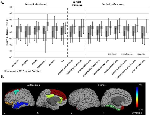
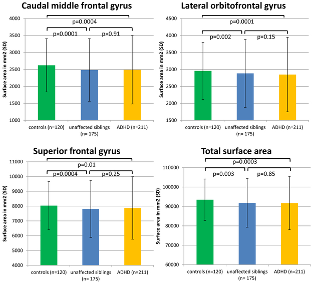
Cortical thickness and surface area (based on the Desikan-Killiany atlas) were compared between case subjects with ADHD (N=2,246) and control subjects (N=1,934) for children, adolescents, and adults separately in ENIGMA-ADHD, a consortium of 36 centers. To assess familial effects on cortical measures, case subjects, unaffected siblings, and control subjects in the NeuroIMAGE study (N=506) were compared. Associations of the attention scale from the Child Behavior Checklist with cortical measures were determined in a pediatric population sample (Generation-R, N=2,707).
In the ENIGMA-ADHD sample, lower surface area values were found in children with ADHD, mainly in frontal, cingulate, and temporal regions; the largest significant effect was for total surface area (Cohen's d=-0.21). Fusiform gyrus and temporal pole cortical thickness was also lower in children with ADHD. Neither surface area nor thickness differences were found in the adolescent or adult groups. Familial effects were seen for surface area in several regions. In an overlapping set of regions, surface area, but not thickness, was associated with attention problems in the Generation-R sample.
Subtle differences in cortical surface area are widespread in children but not adolescents and adults with ADHD, confirming involvement of the frontal cortex and highlighting regions deserving further attention. Notably, the alterations behave like endophenotypes in families and are linked to ADHD symptoms in the population, extending evidence that ADHD behaves as a continuous trait in the population. Future longitudinal studies should clarify individual lifespan trajectories that lead to nonsignificant findings in adolescent and adult groups despite the presence of an ADHD diagnosis.
神经影像学研究表明,注意力缺陷多动障碍(ADHD)患儿和成人的大脑多个区域存在结构改变,尽管重复研究的结果并不一致。作者试图通过大规模研究确定与 ADHD 相关的皮质特征。
ENIGMA-ADHD 研究中,作者对来自 36 个中心的共 2246 例 ADHD 病例和 1934 例对照的儿童、青少年和成人的皮质厚度和皮质表面积(基于 Desikan-Killiany 图谱)进行了比较。为了评估皮质测量值的家族效应,作者对来自 NeuroIMAGE 研究的病例、未受影响的兄弟姐妹和对照(共 506 例)进行了比较。在儿科人群样本(Generation-R,共 2707 例)中,作者确定了儿童行为检查表的注意力量表与皮质测量值的关联。
在 ENIGMA-ADHD 样本中,ADHD 患儿的皮质表面积值较低,主要在额、扣带回和颞叶区域;最大的显著效应是总表面积(Cohen's d=-0.21)。ADHD 患儿的梭状回和颞极皮质厚度也较低。在青少年和成年组中未发现表面积或厚度差异。在几个区域中发现了表面积的家族效应。在 Generation-R 样本的重叠区域中,表面积而非厚度与注意问题相关。
在 ADHD 患儿中,皮质表面积的细微差异广泛存在,但在青少年和成年患者中却不存在,这证实了额叶皮质的受累,并突出了值得进一步关注的区域。值得注意的是,这些改变在家族中表现为表型,并且与人群中的 ADHD 症状相关,进一步证明 ADHD 在人群中是一种连续的特征。未来的纵向研究应阐明导致在青少年和成年组中尽管存在 ADHD 诊断但无显著发现的个体生命轨迹。