Experimental Pathology Unit, Department of Pathology, Faculty of Medicine, McGill University, 3775 University Street, Montreal, Qc, H3A 2B4, Canada.
Present address: Research Acceleration Office, 2001 Campus Delivery, University Services Center, Colorado State University, Fort Collins, CO, 80523, USA.
BMC Cancer. 2019 Apr 24;19(1):376. doi: 10.1186/s12885-019-5587-3.
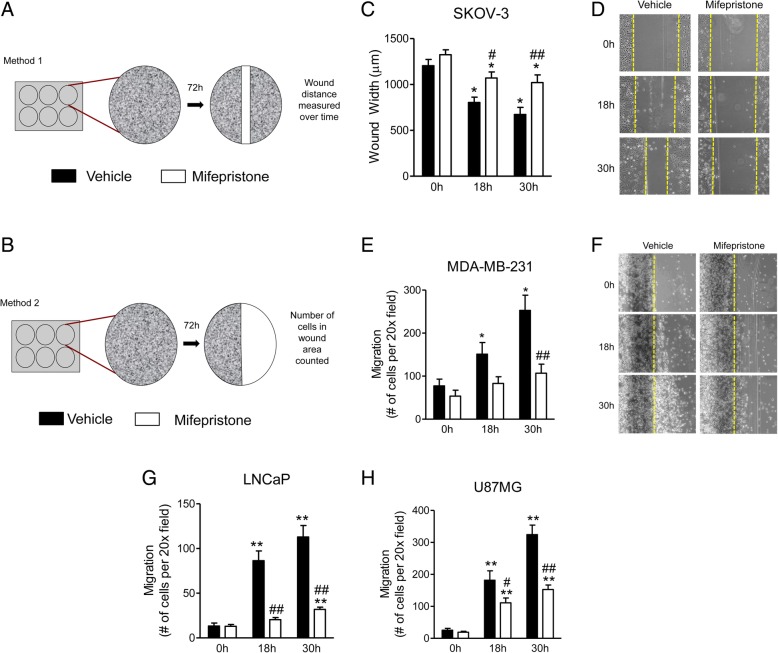
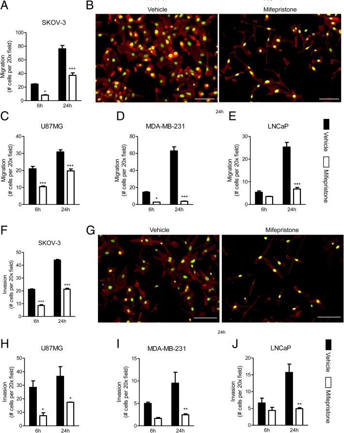
Previous work in our laboratory demonstrated that antiprogestin mifepristone impairs the growth and adhesion of highly metastatic cancer cells, and causes changes in their cellular morphology. In this study, we further assess the anti-metastatic properties of mifepristone, by studying whether cytostatic doses of the drug can inhibit the migration and invasion of various cancer cell lines using a double fluorescence cytochemical labeling approach.
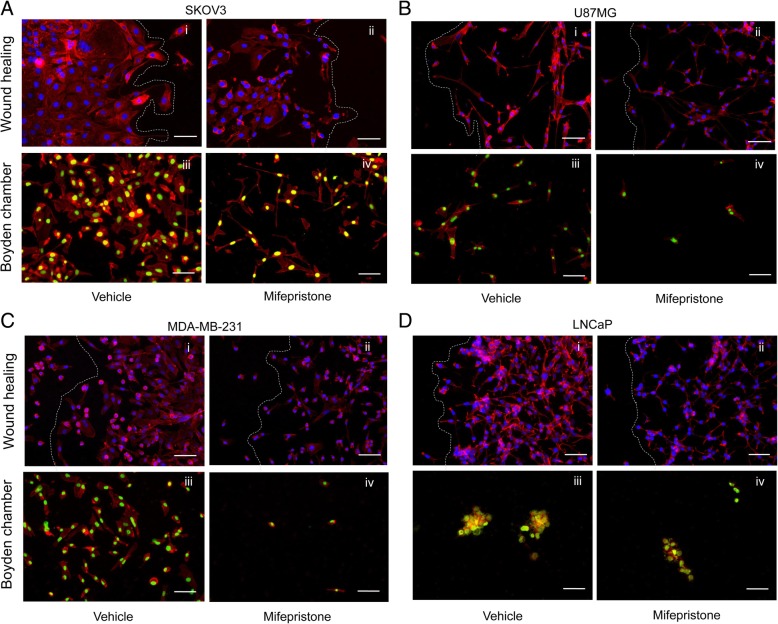
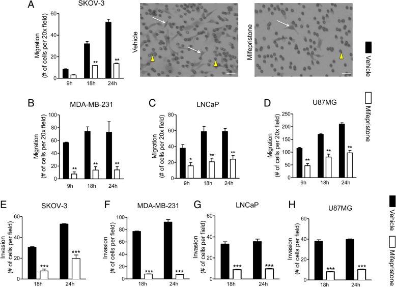
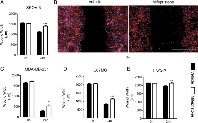
Cell lines representing cancers of the ovary (SKOV-3), breast (MDA-MB-231), glia (U87MG), or prostate (LNCaP) were treated with cytostatic concentrations of mifepristone. Wound healing and Boyden chamber assays were utilized to study cellular migration. To study cellular invasion, the Boyden chamber assay was prepared by adding a layer of extracellular matrix over the polycarbonate membrane. We enhanced the assays with the addition of double fluorescence cytochemical staining for fibrillar actin (F-actin) and DNA to observe the patterns of cytoskeletal distribution and nuclear positioning while cells migrate and invade.
When exposed to cytostatic concentrations of mifepristone, all cancer cells lines demonstrated a decrease in both migration and invasion capacities measured using standard approaches. Double fluorescence cytochemical labeling validated that mifepristone-treated cancer cells exhibit reduced migration and invasion, and allowed to unveil a distinct migration pattern among the different cell lines, different arrays of nuclear localization during migration, and apparent redistribution of F-actin to the nucleus.
This study reports that antiprogestin mifepristone inhibits migration and invasion of highly metastatic cancer cell lines, and that double fluorescence cytochemical labeling increases the value of well-known approaches to study cell movement.
我们实验室之前的工作表明,抗孕激素米非司酮会损害高转移性癌细胞的生长和黏附,并导致其细胞形态发生变化。在这项研究中,我们通过使用双荧光细胞化学标记方法进一步评估米非司酮的抗转移特性,研究细胞毒剂量的药物是否能抑制各种癌细胞系的迁移和侵袭。
用细胞毒浓度的米非司酮处理代表卵巢癌(SKOV-3)、乳腺癌(MDA-MB-231)、神经胶质瘤(U87MG)或前列腺癌(LNCaP)的细胞系。利用划痕愈合和 Boyden 室试验研究细胞迁移。为了研究细胞侵袭,在聚碳酸酯膜上添加一层细胞外基质来制备 Boyden 室试验。通过添加纤维状肌动蛋白(F-actin)和 DNA 的双荧光细胞化学染色,增强了试验,观察细胞迁移和侵袭时细胞骨架分布和核定位的模式。
当暴露于细胞毒浓度的米非司酮时,所有癌细胞系的迁移和侵袭能力均下降,这是使用标准方法测量的。双荧光细胞化学标记证实,米非司酮处理的癌细胞迁移和侵袭能力降低,并且揭示了不同细胞系之间不同的迁移模式、迁移过程中核定位的不同排列以及 F-actin 向核明显再分布。
本研究报告称,抗孕激素米非司酮抑制高转移性癌细胞系的迁移和侵袭,并且双荧光细胞化学标记增加了研究细胞运动的知名方法的价值。