Li Dian, Zhang Dan, Tang Bo, Zhou Yue, Guo Wenhao, Kang Qing, Wang Zhang, Shen Lianju, Wei Guanghui, He Dawei
Chongqing Key Laboratory of Child Urogenital Development and Tissue Engineering, Ministry of Education Key Laboratory of Child Development and Disorders, China International Science and Technology Cooperation Base of Child Development and Critical Disorders, Chongqing Key Laboratory of Pediatrics, Chongqing 400014, China.
Department of Urology, Children's Hospital of Chongqing Medical University, Chongqing 400014, China.
Stem Cells Int. 2019 Mar 19;2019:6935806. doi: 10.1155/2019/6935806. eCollection 2019.
To investigate whether exosomes from human umbilical cord mesenchymal stem cells (hUC-MSCs) can protect against the toxic effects of oxalate and calcium oxalate monohydrate (COM) crystals in human proximal tubular epithelial (HK-2) cells.
Exosomes were isolated from hUC-MSCs, purified by ultracentrifugation, and verified by examination of cell morphology using transmission electron microscopy and the presence of specific biomarkers. HK-2 cells received 1 of 4 treatments: control (cells alone), hUC-MSC exosomes, oxalate+COM, or oxalate+COM and hUC-MSC exosomes. Cell viability was determined using the MTT assay. Oxidative stress was determined by measuring LDH activity and the levels of HO, malondialdehyde (MDA), and reactive oxygen species (ROS). Expressions of N-cadherin, TGF-, and ZO-1 were determined by immunofluorescence. Expressions of epithelial markers, mesenchymal markers, and related signaling pathway proteins were determined by western blotting.
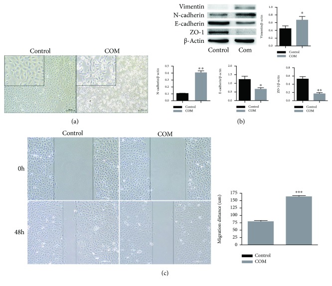
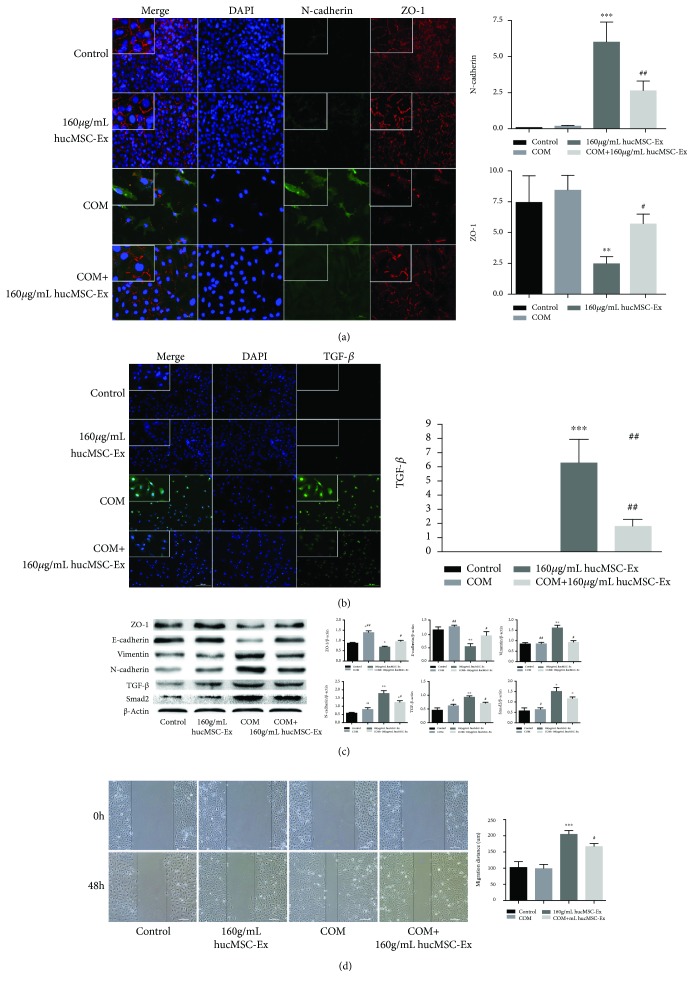
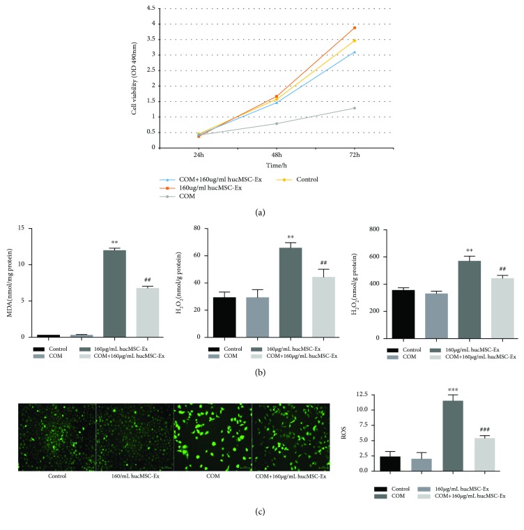
After 48 h, cells in the oxalate+COM group lost their adhesion, appeared long, spindle-shaped, and scattered, and the number of cells had significantly decreased. The oxalate+COM treatment also upregulated TGF- and mesenchymal markers, downregulated epithelial markers, increased the levels of LDH, HO, MDA, and ROS, decreased cell viability, and increased cell migration. The isolated exosomes had double-layer membranes, had hollow, circular, or elliptical shapes, had diameters mostly between 30 and 100 nm, and expressed CD9, CD63, and Alix. Treatment of HK-2 cells with hUC-MSC exosomes reversed or partly reversed all the effects of oxalate+COM.
Exosomes from hUC-MSCs alleviate the oxidative injury and the epithelial-mesenchymal transformation of HK-2 cells that is induced by oxalate+COM.
研究人脐带间充质干细胞(hUC-MSCs)来源的外泌体是否能保护人近端肾小管上皮(HK-2)细胞免受草酸盐和一水合草酸钙(COM)晶体的毒性作用。
从hUC-MSCs中分离外泌体,通过超速离心纯化,并使用透射电子显微镜检查细胞形态和检测特定生物标志物的存在进行验证。HK-2细胞接受4种处理之一:对照(仅细胞)、hUC-MSC外泌体、草酸盐+COM或草酸盐+COM与hUC-MSC外泌体。使用MTT法测定细胞活力。通过测量乳酸脱氢酶(LDH)活性以及过氧化氢(HO)、丙二醛(MDA)和活性氧(ROS)水平来确定氧化应激。通过免疫荧光测定N-钙黏蛋白、转化生长因子-β(TGF-β)和紧密连接蛋白1(ZO-1)的表达。通过蛋白质印迹法测定上皮标志物、间充质标志物和相关信号通路蛋白的表达。
48小时后,草酸盐+COM组的细胞失去黏附性,呈长梭形且分散,细胞数量显著减少。草酸盐+COM处理还上调了TGF-β和间充质标志物,下调了上皮标志物,增加了LDH、HO、MDA和ROS水平,降低了细胞活力,并增加了细胞迁移。分离的外泌体具有双层膜,呈中空、圆形或椭圆形,直径大多在30至100nm之间,并表达CD9、CD63和Alix。用hUC-MSC外泌体处理HK-2细胞可逆转或部分逆转草酸盐+COM的所有作用。
hUC-MSCs来源的外泌体可减轻草酸盐+COM诱导的HK-2细胞氧化损伤和上皮-间充质转化。