Department of Pharmacology and Experimental Therapeutics, Boston University School of Medicine, Boston, Massachusetts, United States of America.
Department of Ophthalmology, Boston University School of Medicine, Boston, Massachusetts, United States of America.
PLoS One. 2019 Apr 24;14(4):e0213422. doi: 10.1371/journal.pone.0213422. eCollection 2019.
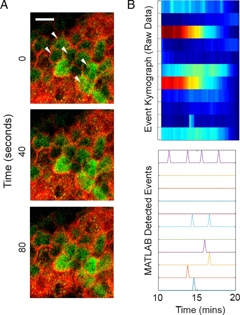
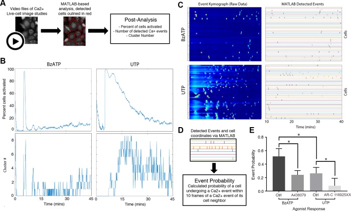
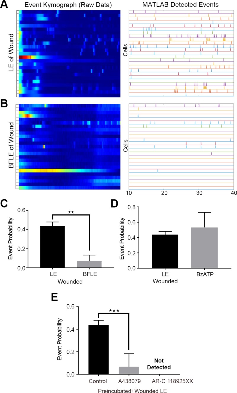
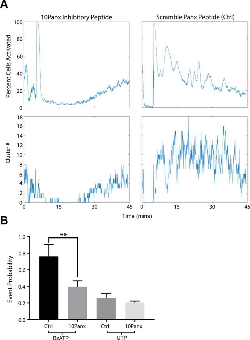
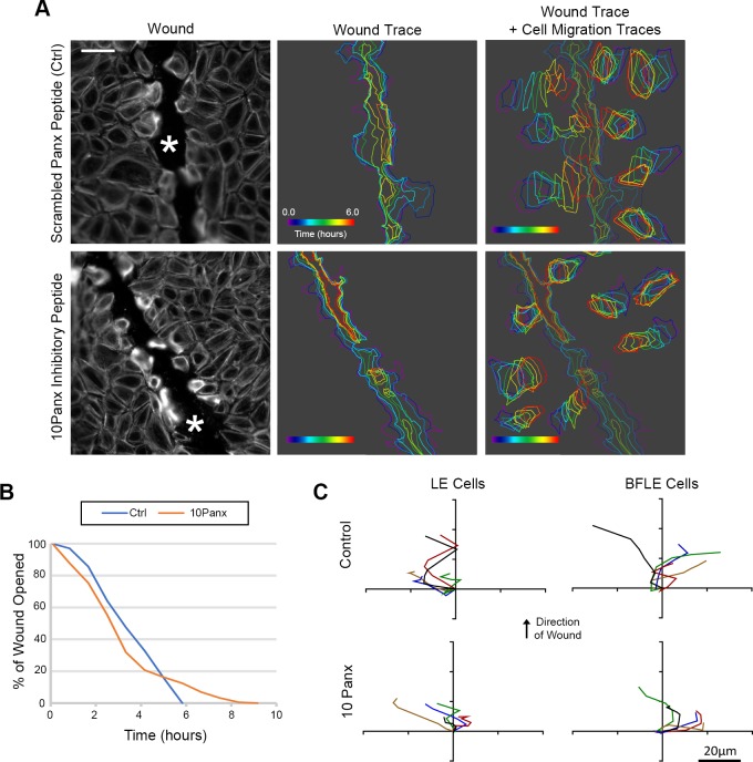
Epithelial wound healing requires the coordination of cells to migrate as a unit over the basement membrane after injury. To understand the process of this coordinated movement, it is critical to study the dynamics of cell-cell communication. We developed a method to characterize the injury-induced sustained Ca2+ mobilizations that travel between cells for periods of time up to several hours. These events of communication are concentrated along the wound edge and are reduced in cells further away from the wound. Our goal was to delineate the role and contribution of these sustained mobilizations and using MATLAB analyses, we determined the probability of cell-cell communication events in both in vitro models and ex vivo organ culture models. We demonstrated that the injury response was complex and represented the activation of a number of receptors. In addition, we found that pannexin channels mediated the cell-cell communication and motility. Furthermore, the sustained Ca2+ mobilizations are associated with changes in cell morphology and motility during wound healing. The results demonstrate that both purinoreceptors and pannexins regulate the sustained Ca2+ mobilization necessary for cell-cell communication in wound healing.
上皮细胞的伤口愈合需要细胞在损伤后协调一致地作为一个整体迁移穿过基底膜。为了理解这个协调运动的过程,研究细胞间通讯的动力学至关重要。我们开发了一种方法来描述损伤诱导的持续 Ca2+动员,这些动员可以在细胞之间传播长达数小时。这些通讯事件集中在伤口边缘,而远离伤口的细胞中的通讯事件则减少。我们的目标是阐明这些持续动员的作用和贡献,并使用 MATLAB 分析,我们在体外模型和离体器官培养模型中确定了细胞间通讯事件的概率。我们证明了损伤反应是复杂的,代表了许多受体的激活。此外,我们发现缝隙连接通道介导了细胞间通讯和运动。此外,持续的 Ca2+动员与伤口愈合过程中细胞形态和运动的变化有关。研究结果表明,嘌呤能受体和缝隙连接通道都调节了伤口愈合中细胞间通讯所必需的持续 Ca2+动员。