Davies Joseph, Riede Philipp, van Langevelde Kirsten, Teh James
Radiology Department, Nuffield Orthopaedic Centre, Windmill Road, Oxford, OX3 7HE, UK.
Nuffield Orthopaedic Centre, Oxford, UK.
Ther Adv Musculoskelet Dis. 2019 Apr 16;11:1759720X19844429. doi: 10.1177/1759720X19844429. eCollection 2019.
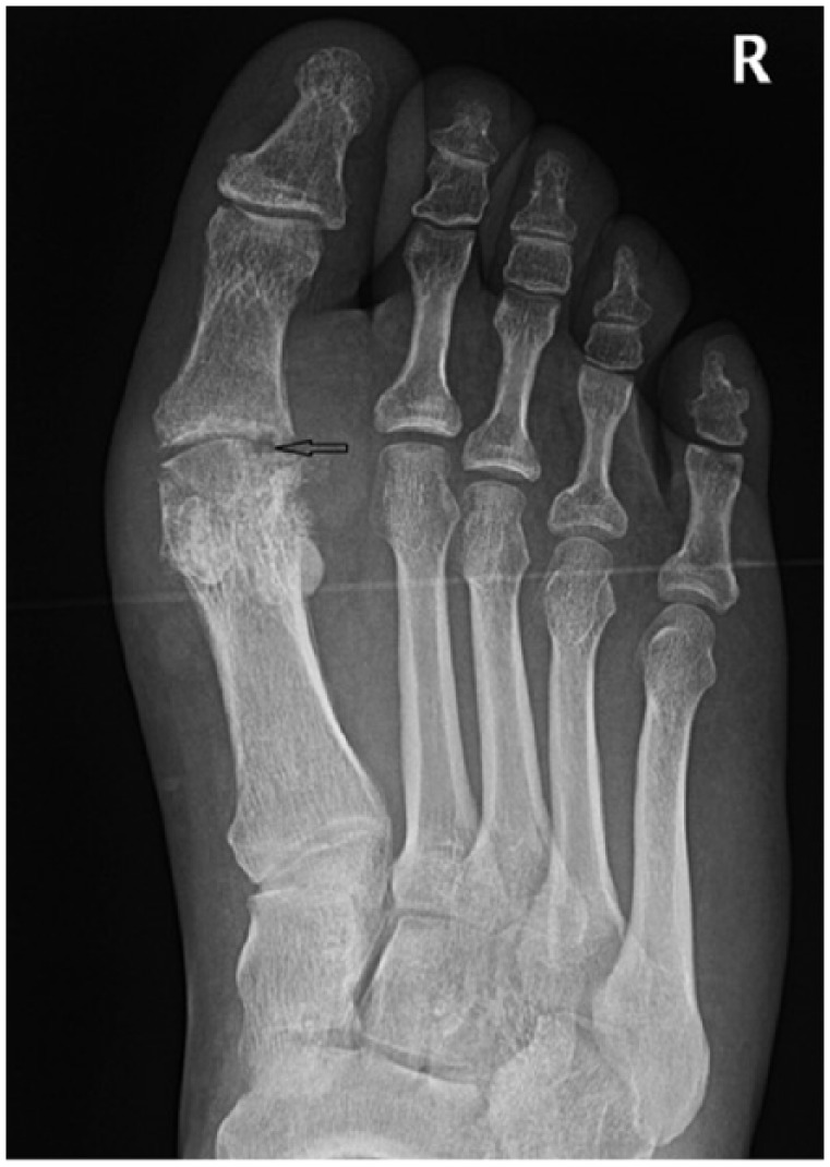
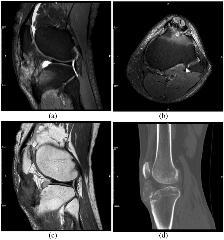
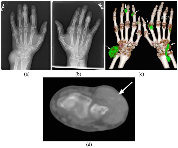
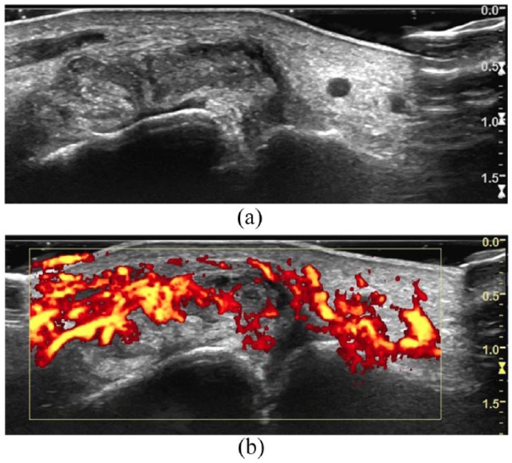
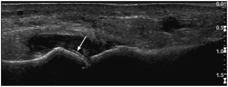
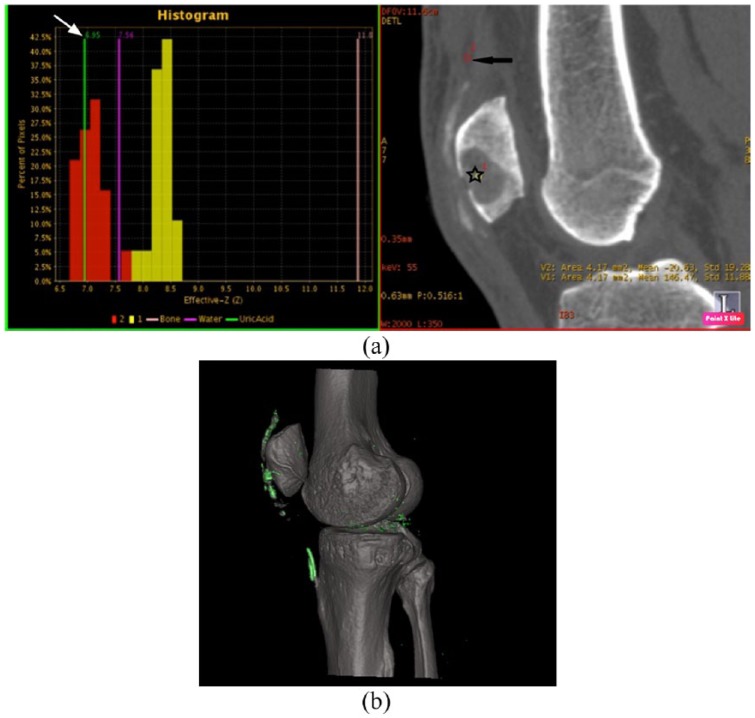
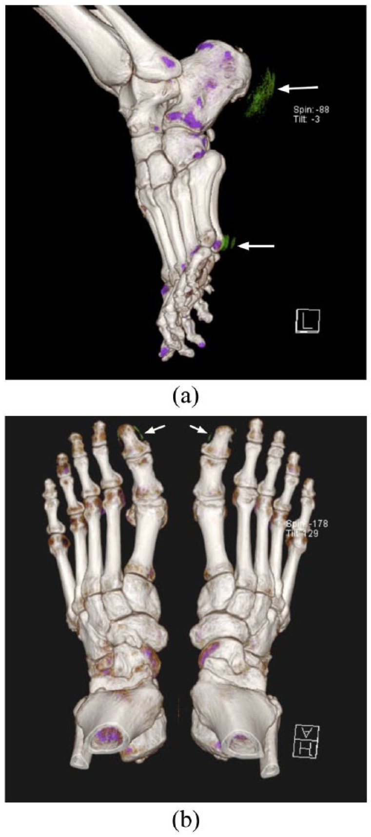
The plain radiographic features of gout are well known; however, the sensitivity of plain radiographs alone for the detection of signs of gout is poor in acute disease. Radiographic abnormalities do not manifest until late in the disease process, after significant joint and soft tissue damage has already occurred. The advent of dual-energy computed tomography (DECT) has enabled the non-invasive diagnosis and quantification of gout by accurately confirming the presence and extent of urate crystals in joints and soft tissues, without the need for painful and often unreliable soft tissue biopsy or joint aspiration. Specific ultrasound findings have been identified and may also be used to aid diagnosis. Both ultrasound and magnetic resonance imaging (MRI) may be used for the measurement of disease extent, monitoring of disease activity or treatment response, although MRI findings are nonspecific. In this article we summarize the imaging findings and diagnostic utility of plain radiographs, ultrasound, DECT, MRI and nuclear medicine studies in the assessment as well as the implications and utility these tools have for measuring disease burden and therapeutic response.
痛风的普通X线特征广为人知;然而,在急性痛风中,仅普通X线片检测痛风体征的敏感性较差。直到疾病后期,在关节和软组织已经发生显著损伤后,X线异常才会显现。双能计算机断层扫描(DECT)的出现,通过准确确认关节和软组织中尿酸盐结晶的存在及范围,实现了痛风的无创诊断和定量分析,无需进行痛苦且通常不可靠的软组织活检或关节穿刺抽吸。已确定了特定的超声表现,其也可用于辅助诊断。超声和磁共振成像(MRI)均可用于测量疾病范围、监测疾病活动或治疗反应,尽管MRI表现不具有特异性。在本文中,我们总结了普通X线片、超声、DECT、MRI和核医学检查在评估中的影像学表现及诊断效用,以及这些工具在测量疾病负担和治疗反应方面的意义和效用。