• 文献检索
  • 文档翻译
  • 深度研究
  • 学术资讯
  • Suppr Zotero 插件Zotero 插件
  • 邀请有礼
  • 套餐&价格
  • 历史记录
应用&插件
Suppr Zotero 插件Zotero 插件浏览器插件Mac 客户端Windows 客户端微信小程序
定价
高级版会员购买积分包购买API积分包
服务
文献检索文档翻译深度研究API 文档MCP 服务
关于我们
关于 Suppr公司介绍联系我们用户协议隐私条款
关注我们

Suppr 超能文献

核心技术专利:CN118964589B侵权必究
粤ICP备2023148730 号-1Suppr @ 2026

文献检索

告别复杂PubMed语法,用中文像聊天一样搜索,搜遍4000万医学文献。AI智能推荐,让科研检索更轻松。

立即免费搜索

文件翻译

保留排版,准确专业,支持PDF/Word/PPT等文件格式,支持 12+语言互译。

免费翻译文档

深度研究

AI帮你快速写综述,25分钟生成高质量综述,智能提取关键信息,辅助科研写作。

立即免费体验

根据胎龄预测胎儿生长速率的胎儿小于胎龄儿。

Prediction of small-for-gestational age by fetal growth rate according to gestational age.

机构信息

Department of Obstetrics and Gynecology, Gangnam Severance Hospital, Institute of Women's Life Medical Science, Yonsei University College of Medicine, Seoul, Korea.

Department of Obstetrics and Gynecology, Severance Hospital, Institute of Women's Life Medical Science, Yonsei University College of Medicine, Seoul, Korea.

出版信息

PLoS One. 2019 Apr 26;14(4):e0215737. doi: 10.1371/journal.pone.0215737. eCollection 2019.

DOI:10.1371/journal.pone.0215737
PMID:31026299
原文链接:https://pmc.ncbi.nlm.nih.gov/articles/PMC6485641/
Abstract

BACKGROUND

Small-for-gestational age (SGA) infants should be identified before birth because of an increased risk of adverse perinatal outcomes. The objective of this study was to assess the impact of fetal growth rate by gestational age on the prediction of SGA and to identify the optimal time to initiate intensive fetal monitoring to detect SGA in low-risk women. We also sought to determine which the ultrasonographic parameters that contribute substantially to the birthweight determination.

METHODS

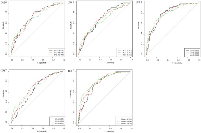
This was a retrospective study of 442 healthy pregnant women with singleton pregnancies. There were 328 adequate-for-gestational age (AGA) neonates and 114 SGA infants delivered between 37+0 and 41+6 weeks of gestation. We compared the biparietal diameters (BPD), head circumferences (HC), abdominal circumferences (AC), femur lengths (FL), and estimated fetal weights (EFW) obtained on each ultrasound to determine which of these parameters was the best indicator of SGA. We created receiver operating characteristic curves, calculated the areas under the curves (AUCs), and analyzed the data using multivariable logistic regressions to assess the ultrasound screening performances and identify the best predictive factor.

RESULTS

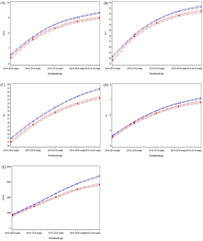
Among the four ultrasonographic parameters, the AC measurement between 24+028+6 weeks achieved a sensitivity of 79.5% and a specificity of 71.7%, with an AUC of 0.806 in the prediction of SGA. AC showed consistently higher AUCs above 0.8 with 6480% sensitivities as gestational age progressed. EFW measurements from 33+035+6 gestational weeks achieved a sensitivity of 60.6% and a specificity of 87.6%, with an AUC of 0.826. In a conditional growth model developed from the linear mixed regression, the value differences between AC and EFW in the SGA and AGA groups became even more pronounced after 33+035+6 weeks.

CONCLUSION

Healthy low-risk women with a low fetal AC after 24 weeks' gestation need to be monitored carefully for fetal growth to identify SGA infants with a risk for adverse perinatal outcomes.

摘要

背景

由于围产期不良结局的风险增加,应在出生前识别出小于胎龄儿(SGA)。本研究的目的是评估胎龄相关的胎儿生长速度对 SGA 预测的影响,并确定在低危孕妇中开始强化胎儿监测以发现 SGA 的最佳时间。我们还试图确定哪些超声参数对确定出生体重有重要贡献。

方法

这是一项回顾性研究,纳入了 442 名单胎妊娠的健康孕妇。其中 328 名胎儿大小与胎龄相符(AGA),114 名 SGA 婴儿在 37+0 至 41+6 周之间分娩。我们比较了每个超声检查中获得的双顶径(BPD)、头围(HC)、腹围(AC)、股骨长(FL)和估计胎儿体重(EFW),以确定这些参数中哪一个是 SGA 的最佳指标。我们绘制了受试者工作特征曲线,计算了曲线下面积(AUC),并使用多变量逻辑回归分析数据,以评估超声筛查性能并确定最佳预测因素。

结果

在四个超声参数中,24+028+6 周的 AC 测量在预测 SGA 方面具有 79.5%的敏感性和 71.7%的特异性,AUC 为 0.806。随着胎龄的增加,AC 的 AUC 始终高于 0.8,其敏感性为 64%80%。33+035+6 周的 EFW 测量具有 60.6%的敏感性和 87.6%的特异性,AUC 为 0.826。在从线性混合回归中建立的条件生长模型中,在 33+035+6 周之后,SGA 和 AGA 组中 AC 和 EFW 之间的差值变得更加明显。

结论

在妊娠 24 周后,胎龄较小的健康低危孕妇需要仔细监测胎儿生长情况,以识别有不良围产期结局风险的 SGA 婴儿。

https://cdn.ncbi.nlm.nih.gov/pmc/blobs/141b/6485641/5a3f5e6d87d0/pone.0215737.g002.jpg
https://cdn.ncbi.nlm.nih.gov/pmc/blobs/141b/6485641/a74738400664/pone.0215737.g001.jpg
https://cdn.ncbi.nlm.nih.gov/pmc/blobs/141b/6485641/5a3f5e6d87d0/pone.0215737.g002.jpg
https://cdn.ncbi.nlm.nih.gov/pmc/blobs/141b/6485641/a74738400664/pone.0215737.g001.jpg
https://cdn.ncbi.nlm.nih.gov/pmc/blobs/141b/6485641/5a3f5e6d87d0/pone.0215737.g002.jpg

相似文献

1
Prediction of small-for-gestational age by fetal growth rate according to gestational age.根据胎龄预测胎儿生长速率的胎儿小于胎龄儿。
PLoS One. 2019 Apr 26;14(4):e0215737. doi: 10.1371/journal.pone.0215737. eCollection 2019.
2
Prediction of small-for-gestational-age neonates at 35-37 weeks' gestation: contribution of maternal factors and growth velocity between 32 and 36 weeks.预测 35-37 孕周的小于胎龄儿:母亲因素的影响和 32-36 孕周间的生长速度。
Ultrasound Obstet Gynecol. 2019 May;53(5):630-637. doi: 10.1002/uog.20267. Epub 2019 Apr 8.
3
Routine ultrasound at 32 vs 36 weeks' gestation: prediction of small-for-gestational-age neonates.32 周与 36 周常规超声检查:预测小于胎龄儿。
Ultrasound Obstet Gynecol. 2019 Jun;53(6):761-768. doi: 10.1002/uog.20258. Epub 2019 Apr 30.
4
Two-stage approach for prediction of small-for-gestational-age neonate and adverse perinatal outcome by routine ultrasound examination at 35-37 weeks' gestation.在 35-37 孕周的常规超声检查中预测小于胎龄儿和不良围产结局的两阶段方法。
Ultrasound Obstet Gynecol. 2019 Oct;54(4):484-491. doi: 10.1002/uog.20391. Epub 2019 Aug 27.
5
Prediction of small-for-gestational-age neonates at 35-37 weeks' gestation: contribution of maternal factors and growth velocity between 20 and 36 weeks.预测 35-37 孕周的小于胎龄儿:母亲因素和 20-36 孕周间生长速度的作用。
Ultrasound Obstet Gynecol. 2019 Apr;53(4):488-495. doi: 10.1002/uog.20243.
6
Single and Serial Fetal Biometry to Detect Preterm and Term Small- and Large-for-Gestational-Age Neonates: A Longitudinal Cohort Study.单胎及多胎胎儿生物测量用于检测早产及足月的小于胎龄儿和大于胎龄儿:一项纵向队列研究。
PLoS One. 2016 Nov 1;11(11):e0164161. doi: 10.1371/journal.pone.0164161. eCollection 2016.
7
Prediction of small-for-gestational-age neonates at 33-39 weeks' gestation in China: logistic regression modeling of the contributions of second- and third-trimester ultrasound data and maternal factors.中国 33-39 孕周小胎龄儿的预测:二、三孕期超声数据和母体因素的逻辑回归模型贡献。
BMC Pregnancy Childbirth. 2022 Aug 25;22(1):661. doi: 10.1186/s12884-022-04991-7.
8
Third trimester ultrasound estimated fetal weight for increasing prenatal prediction of small-for-gestational age newborns in low-risk pregnant women.孕晚期超声评估胎儿体重,以提高对低风险孕妇中小于胎龄新生儿的产前预测。
J Matern Fetal Neonatal Med. 2022 Dec;35(25):6721-6726. doi: 10.1080/14767058.2021.1920915. Epub 2021 May 23.
9
Prediction of large-for-gestational-age neonate by routine third-trimester ultrasound.通过常规孕晚期超声预测大于胎龄儿。
Ultrasound Obstet Gynecol. 2019 Sep;54(3):326-333. doi: 10.1002/uog.20377. Epub 2019 Jul 23.
10
Comparing the relation between ultrasound-estimated fetal weight and birthweight in cohort of small-for-gestational-age fetuses.比较小胎龄儿队列中超声估计胎儿体重与出生体重的关系。
Acta Obstet Gynecol Scand. 2019 Nov;98(11):1435-1441. doi: 10.1111/aogs.13645. Epub 2019 Jun 6.

引用本文的文献

1
Maternal BMI and Diagnostic Accuracy of Using Estimated Fetal Growth to Predict Abnormal Birthweight: Results from NICHD Fetal Growth Studies.母亲体重指数与利用估计胎儿生长预测异常出生体重的诊断准确性:来自美国国立儿童健康与人类发展研究所胎儿生长研究的结果
Diagnostics (Basel). 2025 May 31;15(11):1398. doi: 10.3390/diagnostics15111398.
2
Prognostic prediction models for adverse birth outcomes: A systematic review.预测不良出生结局的预后预测模型:系统评价。
J Glob Health. 2024 Oct 25;14:04214. doi: 10.7189/jogh.14.04214.
3
Prediction of small-for-gestational-age neonates at 33-39 weeks' gestation in China: logistic regression modeling of the contributions of second- and third-trimester ultrasound data and maternal factors.

本文引用的文献

1
The Hadlock Method Is Superior to Newer Methods for the Prediction of the Birth Weight Percentile.哈德洛克方法在预测出生体重百分位数方面优于新方法。
J Ultrasound Med. 2019 Mar;38(3):587-596. doi: 10.1002/jum.14725. Epub 2018 Sep 23.
2
Factors related to low birth rate among married women in Korea.韩国已婚女性低生育率相关因素。
PLoS One. 2018 Mar 20;13(3):e0194597. doi: 10.1371/journal.pone.0194597. eCollection 2018.
3
Customized growth charts: rationale, validation and clinical benefits.定制生长图表:原理、验证和临床获益。
中国 33-39 孕周小胎龄儿的预测:二、三孕期超声数据和母体因素的逻辑回归模型贡献。
BMC Pregnancy Childbirth. 2022 Aug 25;22(1):661. doi: 10.1186/s12884-022-04991-7.
4
Estimation of multiple ordered ROC curves using placement values.使用定位值估计多个有序 ROC 曲线。
Stat Methods Med Res. 2022 Aug;31(8):1470-1483. doi: 10.1177/09622802221094940. Epub 2022 Apr 21.
5
Alterations of brain metrics in fetuses of women with polycystic ovary syndrome : a retrospective study based on fetal magnetic resonance imaging.多囊卵巢综合征女性胎儿脑参数改变:基于胎儿磁共振成像的回顾性研究。
BMC Pregnancy Childbirth. 2021 Aug 14;21(1):557. doi: 10.1186/s12884-021-04015-w.
6
Fetal growth, fetal development, and placental features in women with polycystic ovary syndrome: analysis based on fetal and placental magnetic resonance imaging.多囊卵巢综合征女性的胎儿生长、胎儿发育和胎盘特征:基于胎儿和胎盘磁共振成像的分析。
J Zhejiang Univ Sci B. 2020;21(12):977-989. doi: 10.1631/jzus.B2000350.
Am J Obstet Gynecol. 2018 Feb;218(2S):S609-S618. doi: 10.1016/j.ajog.2017.12.011.
4
Fetal growth standards: the NICHD fetal growth study approach in context with INTERGROWTH-21st and the World Health Organization Multicentre Growth Reference Study.胎儿生长标准:NICHD 胎儿生长研究方法与 INTERGROWTH-21st 和世界卫生组织多中心生长参考研究的关系。
Am J Obstet Gynecol. 2018 Feb;218(2S):S641-S655.e28. doi: 10.1016/j.ajog.2017.11.593. Epub 2017 Dec 22.
5
The World Health Organization Fetal Growth Charts: A Multinational Longitudinal Study of Ultrasound Biometric Measurements and Estimated Fetal Weight.世界卫生组织胎儿生长图表:超声生物测量与估计胎儿体重的多国纵向研究
PLoS Med. 2017 Jan 24;14(1):e1002220. doi: 10.1371/journal.pmed.1002220. eCollection 2017 Jan.
6
International estimated fetal weight standards of the INTERGROWTH-21 Project.INTERGROWTH-21项目的国际胎儿体重估计标准。
Ultrasound Obstet Gynecol. 2017 Apr;49(4):478-486. doi: 10.1002/uog.17347. Epub 2017 Mar 5.
7
Single and Serial Fetal Biometry to Detect Preterm and Term Small- and Large-for-Gestational-Age Neonates: A Longitudinal Cohort Study.单胎及多胎胎儿生物测量用于检测早产及足月的小于胎龄儿和大于胎龄儿:一项纵向队列研究。
PLoS One. 2016 Nov 1;11(11):e0164161. doi: 10.1371/journal.pone.0164161. eCollection 2016.
8
Prediction of small-for-gestational-age neonate by third-trimester fetal biometry and impact of ultrasound-delivery interval.孕晚期胎儿生物测量对小于胎龄儿的预测及超声检查与分娩间隔的影响
Ultrasound Obstet Gynecol. 2017 Mar;49(3):372-378. doi: 10.1002/uog.15959.
9
Screening for fetal growth restriction with universal third trimester ultrasonography in nulliparous women in the Pregnancy Outcome Prediction (POP) study: a prospective cohort study.妊娠结局预测(POP)研究中对未生育女性进行孕晚期常规超声检查以筛查胎儿生长受限:一项前瞻性队列研究。
Lancet. 2015 Nov 21;386(10008):2089-2097. doi: 10.1016/S0140-6736(15)00131-2. Epub 2015 Sep 7.
10
Ultrasound screening for fetal growth restriction at 36 vs 32 weeks' gestation: a randomized trial (ROUTE).孕36周与32周超声筛查胎儿生长受限:一项随机试验(ROUTE)
Ultrasound Obstet Gynecol. 2015 Oct;46(4):391-7. doi: 10.1002/uog.14915.