Li Na, Zhang Fu-Bin, Li Bing-Jie, Wang Rui-Tao
Department of Internal Medicine, Harbin Medical University Cancer Hospital, Harbin Medical University, Harbin, Heilongjiang, China. Email:
Department of Gynecology, Harbin Medical University Cancer Hospital, Harbin Medical University, Harbin, Heilongjiang,China.
Asian Pac J Cancer Prev. 2019 Apr 29;20(4):1025-1029. doi: 10.31557/APJCP.2019.20.4.1025.
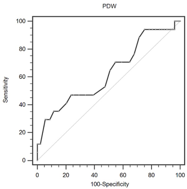
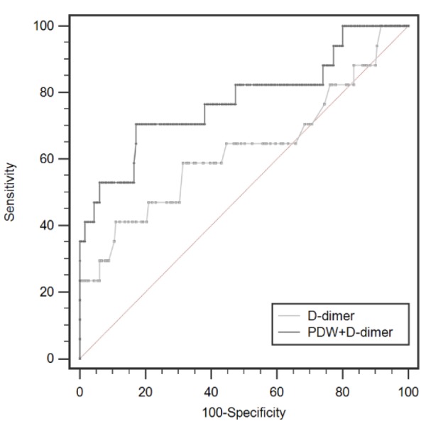
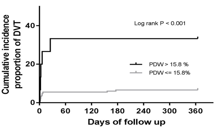
Background: Deep venous thrombosis (DVT) is associated with severe morbidity and mortality in cancer. Platelet distribution width (PDW), a platelet index, indicates variation in platelet size. We aimed to investigate whether the combination of D-dimer and PDW could have a better performance in predicting DVT in patients with cervical carcinoma. Materials and Methods: In 198 consecutive cervical carcinoma patients without preoperative DVT, preoperative D-dimer and PDW levels were measured. Compression ultrasonography was performed in all cervical carcinoma patients before surgery, as well as one month, three months, six months, and 12 months. Results: During a median period of 12 months, 17 of the 198 patients (8.6 %) developed DVT. PDW levels were reduced and D-dimer levels were increased in patients with DVT events compared to those without DVT. Multivariate Cox analysis revealed that both PDW and D-dimer were independent predictors for DVT events. The area under the ROC curve was 0.628 (95% CI: 0.556 to 0.695, p=0.142) when D-dimer was used alone, whereas it increased to 0.777 (95% CI: 0.712 to 0.833, p<0.011) with the addition of PDW. Incorporation of PDW into the D-dimer model significantly improved the predictive value. Conclusions: The combination of preoperative D-dimer and PDW improves the predictive power of postoperative DVT risk in patients with cervical carcinoma.
深静脉血栓形成(DVT)与癌症患者的严重发病率和死亡率相关。血小板分布宽度(PDW)是一种血小板指标,可反映血小板大小的变化。我们旨在研究D-二聚体和PDW的联合检测在预测宫颈癌患者DVT方面是否具有更好的性能。材料与方法:对198例术前无DVT的宫颈癌患者进行术前D-二聚体和PDW水平检测。所有宫颈癌患者在手术前以及术后1个月、3个月、6个月和12个月均进行了超声检查。结果:在中位随访期12个月内,198例患者中有17例(8.6%)发生了DVT。与未发生DVT的患者相比,发生DVT事件的患者PDW水平降低,D-二聚体水平升高。多因素Cox分析显示,PDW和D-二聚体均为DVT事件的独立预测因素。单独使用D-二聚体时,ROC曲线下面积为0.628(95%CI:0.556至0.695,p=0.142),而加入PDW后,该面积增加至0.777(95%CI:0.712至0.833,p<0.011)。将PDW纳入D-二聚体模型可显著提高预测价值。结论:术前D-二聚体和PDW的联合检测可提高宫颈癌患者术后DVT风险的预测能力。