Univ Rennes, CHU Rennes, Laboratoire de Biologie de la Reproduction -CECOS, Inserm, EHESP, Irset (Institut de recherche en santé, environnement et travail) - UMR_S 1085, Rennes F-35000, France.
Sechenov Institute of Evolutionary Physiology and Biochemistry, Russian Academy of Sciences, 194223 St-Petersburg, Russia.
Asian J Androl. 2019 Nov-Dec;21(6):570-576. doi: 10.4103/aja.aja_12_19.
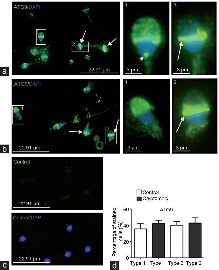
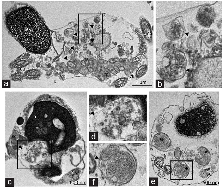
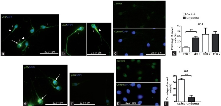
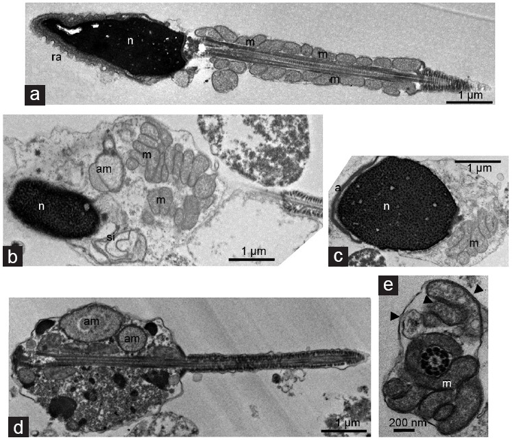
Autophagy is involved in spermatogenesis by regulating germ cell maturation. This catabolic process increases with hyperthermic conditions to prevent the accumulation of damaged organelles. Cryptorchidism is associated with impairment of germ cell maturation revealed by the presence of immature forms of sperm cells in ejaculates. The aim of the present study was to evaluate the status of autophagy in sperm cells from cryptorchid patients. Semen samples of cryptorchid patients and normozoospermic controls were analyzed by immunocytochemistry and electron microscopy. Autophagy proteins, autophagy-related protein 9 (ATG9) and microtubule-associated protein, 1A/1B-light chain 3 (LC3) were localized by immunocytochemistry on the acrosome and on the equatorial segment of sperm cells. LC3 was also detected in the midpiece of cryptorchid sperm tail. Autophagy substrate p62 protein was present in the acrosome and in the postequatorial segment of sperm in control samples, but not in the cryptorchid ones. Transmission electron microscopy revealed double-membrane-limited autophagosomes in postequatorial part of spermatozoa head and midpiece in cryptorchid samples. Partly degraded mitochondria were frequently discerned in autophagic vacuoles. In conclusion, autophagy is increased in sperm cells from patients with cryptorchid history comparatively to control. Our work provides insights into the role of autophagy in the maturation and survival of human male gametes in pathological conditions. Thus, regulating autophagy could represent a potential way to improve sperm quality in cryptorchid men.
自噬通过调节精母细胞成熟参与精子发生。这个分解代谢过程随着高温条件的增加而增加,以防止受损细胞器的积累。隐睾症与精母细胞成熟受损有关,表现在精液中存在不成熟的精子细胞。本研究旨在评估隐睾症患者精子细胞中自噬的状态。通过免疫细胞化学和电子显微镜分析隐睾症患者和正常精子症对照者的精液样本。自噬蛋白、自噬相关蛋白 9 (ATG9) 和微管相关蛋白 1A/1B-轻链 3 (LC3) 通过免疫细胞化学在精子细胞的顶体和赤道段上定位。LC3 也在隐睾症精子尾部的中段被检测到。自噬底物 p62 蛋白存在于对照组精子的顶体和赤道段后段,但不存在于隐睾症患者的精子中。透射电子显微镜显示,隐睾症精子头部和中段的赤道段后部分别有双层膜自噬体。自噬小体中经常可以识别到部分降解的线粒体。总之,与对照组相比,隐睾症患者精子细胞中的自噬增加。我们的工作为自噬在病理条件下人类精子成熟和存活中的作用提供了新的见解。因此,调节自噬可能是提高隐睾症男性精子质量的一种潜在方法。