Section of Radiology, Department of Surgical Sciences, Uppsala University, Uppsala, Sweden.
Antaros Medical, Mölndal, Sweden.
Magn Reson Med. 2019 Sep;82(3):1177-1186. doi: 10.1002/mrm.27786. Epub 2019 Apr 29.
To perform and evaluate water-fat signal separation of whole-body gradient echo scans using convolutional neural networks.
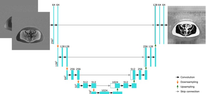
Whole-body gradient echo scans of 240 subjects, each consisting of 5 bipolar echoes, were used. Reference fat fraction maps were created using a conventional method. Convolutional neural networks, more specifically 2D U-nets, were trained using 5-fold cross-validation with 1 or several echoes as input, using the squared difference between the output and the reference fat fraction maps as the loss function. The outputs of the networks were assessed by the loss function, measured liver fat fractions, and visually. Training was performed using a graphics processing unit (GPU). Inference was performed using the GPU as well as a central processing unit (CPU).
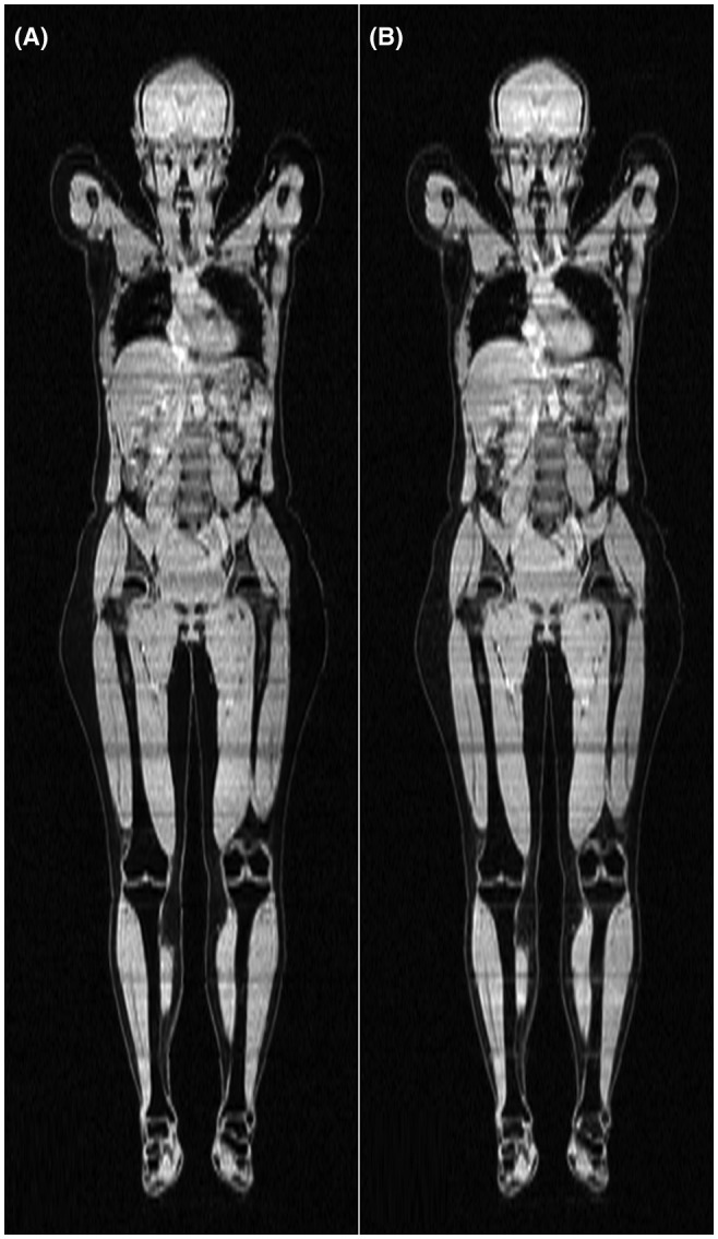
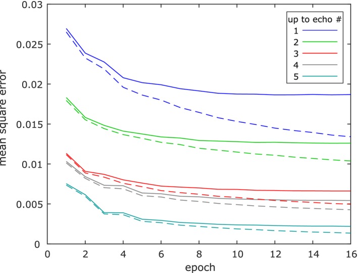
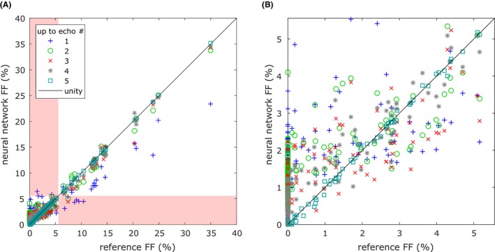
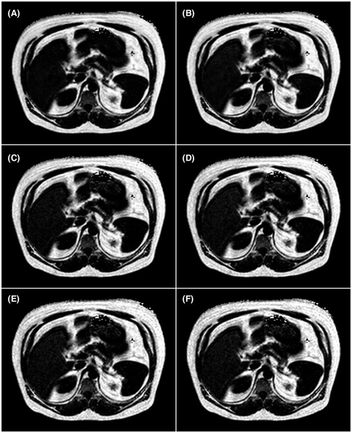
The loss curves indicated convergence, and the final loss of the validation data decreased when using more echoes as input. The liver fat fractions could be estimated using only 1 echo, but results were improved by use of more echoes. Visual assessment found the quality of the outputs of the networks to be similar to the reference even when using only 1 echo, with slight improvements when using more echoes. Training a network took at most 28.6 h. Inference time of a whole-body scan took at most 3.7 s using the GPU and 5.8 min using the CPU.
It is possible to perform water-fat signal separation of whole-body gradient echo scans using convolutional neural networks. Separation was possible using only 1 echo, although using more echoes improved the results.
使用卷积神经网络对全身梯度回波扫描进行水脂信号分离并进行评估。
使用了 240 名受试者的全身梯度回波扫描,每个扫描由 5 个双极回波组成。参考脂肪分数图是使用传统方法创建的。卷积神经网络,更具体地说是 2D U-Net,使用 5 折交叉验证,以 1 个或多个回波作为输入,使用输出与参考脂肪分数图之间的平方差作为损失函数进行训练。网络的输出通过损失函数、测量的肝脂肪分数和视觉评估来评估。训练使用图形处理单元 (GPU) 进行。推断使用 GPU 以及中央处理单元 (CPU) 进行。
损失曲线表明已经收敛,并且当使用更多的回波作为输入时,验证数据的最终损失会降低。仅使用 1 个回波就可以估计肝脂肪分数,但使用更多的回波可以提高结果。视觉评估发现,即使仅使用 1 个回波,网络输出的质量也与参考图相似,使用更多的回波时会有一些改进。训练一个网络最多需要 28.6 小时。使用 GPU 进行全身扫描的推断时间最多为 3.7 秒,使用 CPU 则需要 5.8 分钟。
使用卷积神经网络对全身梯度回波扫描进行水脂信号分离是可行的。虽然使用更多的回波可以提高结果,但仅使用 1 个回波也可以进行分离。