Department of Laboratory Medicine, Seoul National University College of Medicine, Seoul, Republic of Korea.
Cancer Research Institute, Seoul National University College of Medicine, Seoul, Republic of Korea.
PLoS One. 2019 Apr 29;14(4):e0216055. doi: 10.1371/journal.pone.0216055. eCollection 2019.
Neutrophils can generate extracellular net-like structures by releasing their DNA-histone complexes and antimicrobial peptides, which is called neutrophil extracellular traps (NETs). Various stimuli can induce NET formation. In particular, neutrophils and NET formation are abundant in tumor tissue. This study investigated how cancer cells induce NET formation and whether this NET formation promotes plasma thrombin generation and cancer progression.
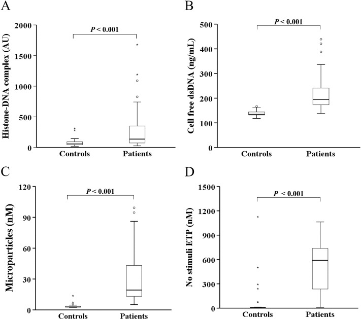
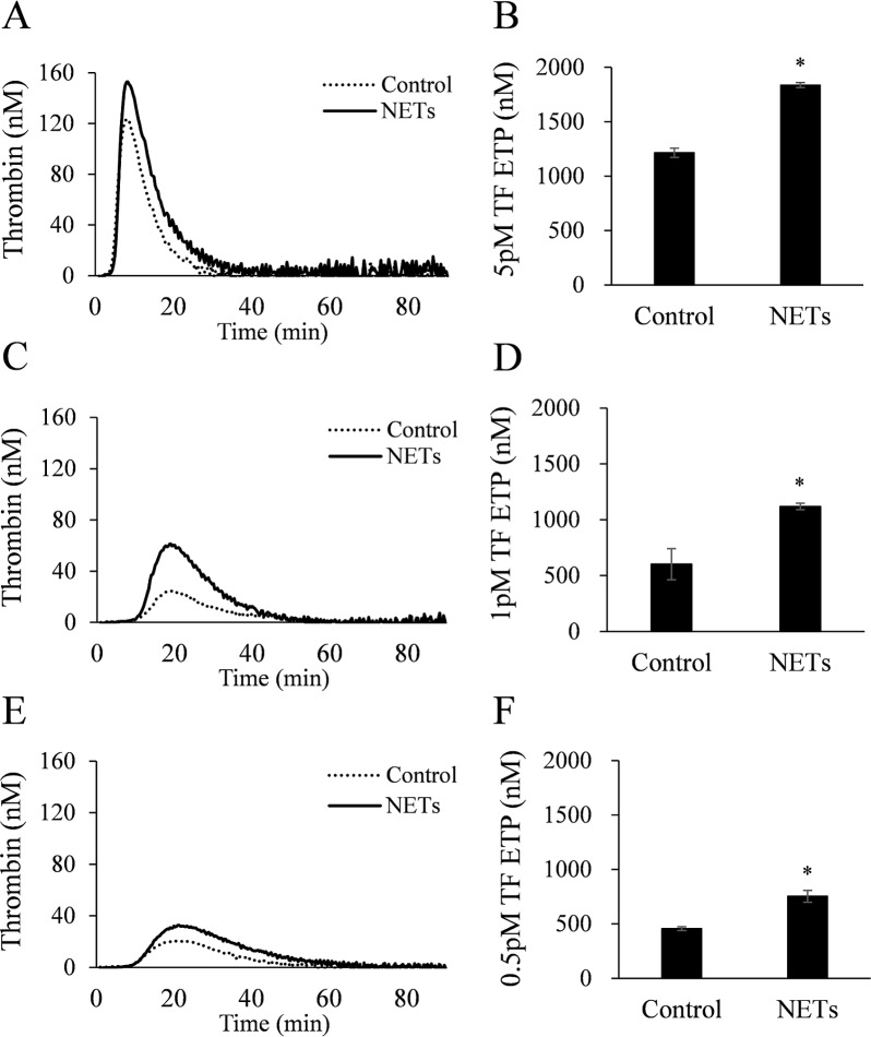
Induction of NET formation by a pancreatic cancer cell line (AsPC-1) was assessed by measuring the histone-DNA complex level. The endogenous thrombin potential (ETP) was measured by thrombin generation assay. In vitro migration, invasion, and tubule formation assays were performed. The circulating levels of NET markers and hypercoagulability markers were assessed in 62 patients with pancreatobiliary malignancy and 30 healthy controls.
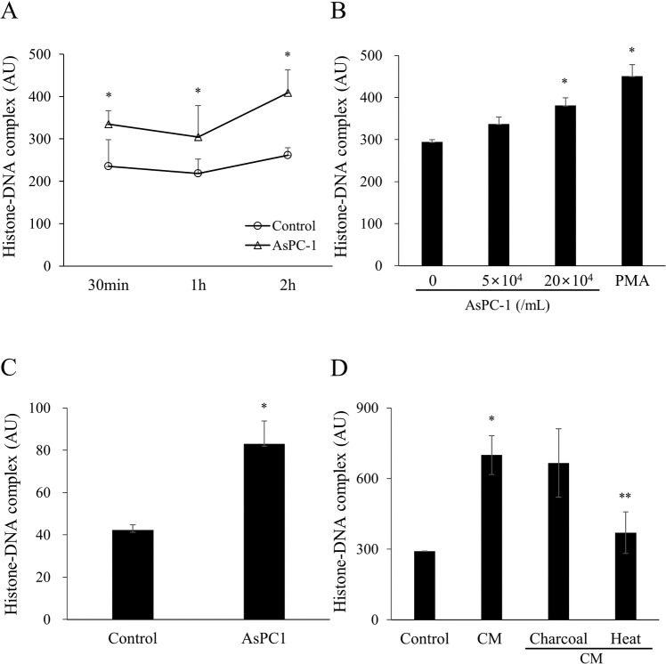
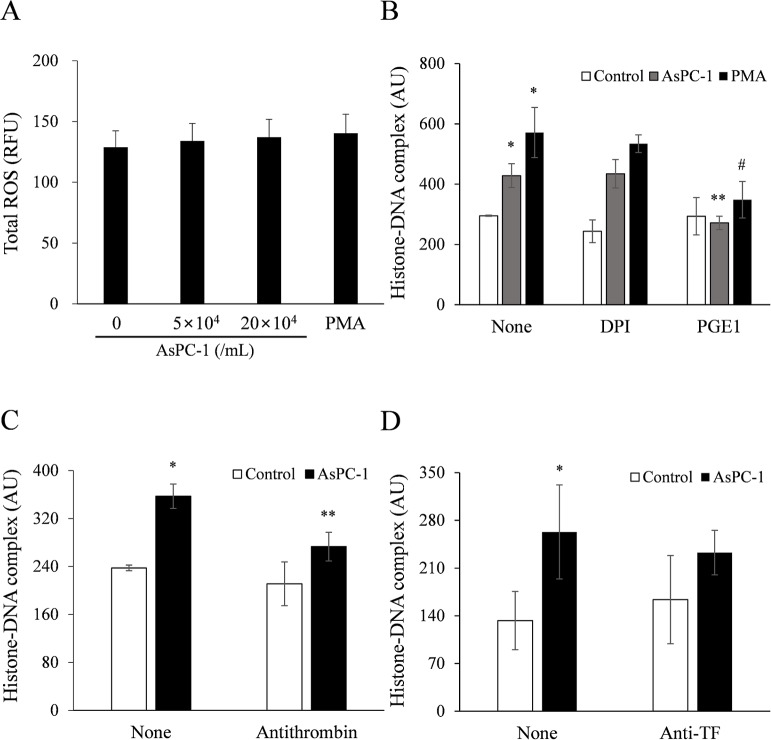
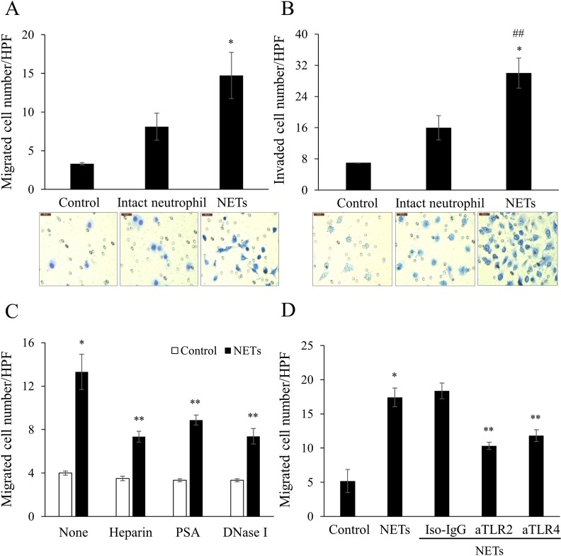
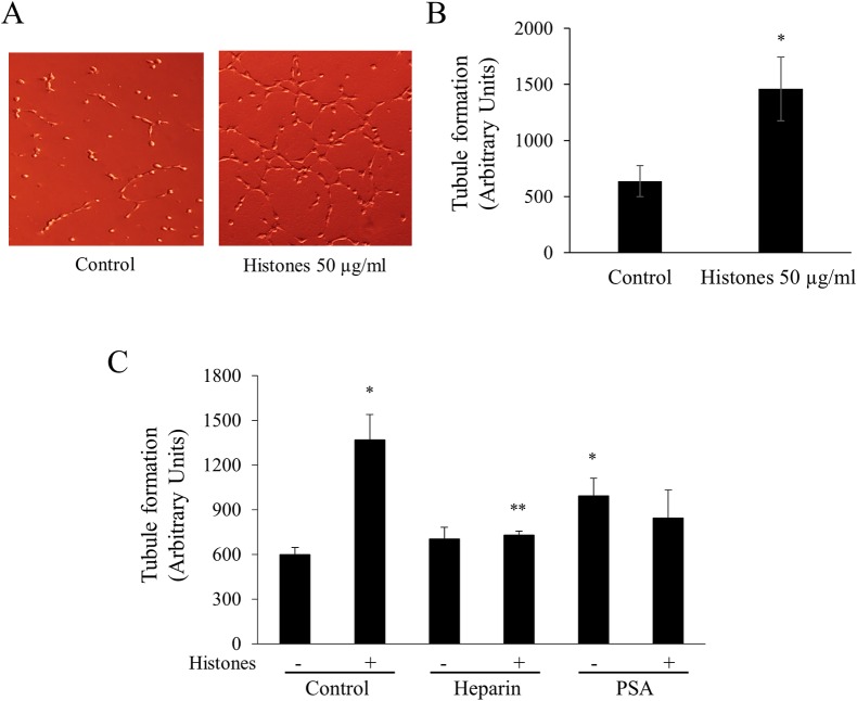
AsPC-1 significantly induced NET formation in a dose-dependent manner. Conditioned medium (CM) from AsPC-1 also induced NETs. Interestingly, NET-formation was abolished by heat-inactivated CM, but not by lipid-extracted CM, suggesting an important role of protein components. A reactive oxygen species inhibitor did not inhibit cancer cell-induced NET formation, but prostaglandin E1 (PGE1, cyclic adenosine monophosphate inducer) and antithrombin did. NETs significantly increased ETP of normal plasma. Of note, NETs promoted cancer cell migration and invasion as well as angiogenesis, which were inhibited by histone-binding agents (heparin, polysialic acid), a DNA-degrading enzyme, and Toll-like receptor neutralizing antibodies. In patients with pancreatobiliary malignancy, elevated NET markers correlated well with hypercoagulability makers.
Our findings indicate that cancer cell-induced NET formation enhances both hypercoagulability and cancer progression and suggest that inhibitors of NET formation such as PGE1 and antithrombin can be potential therapeutics to reduce both hypercoagulability and cancer progression.
中性粒细胞可以通过释放其 DNA-组蛋白复合物和抗菌肽来生成细胞外网状结构,这被称为中性粒细胞胞外诱捕网(NETs)。各种刺激可以诱导 NET 的形成。特别是,在肿瘤组织中,中性粒细胞和 NET 的形成很丰富。本研究探讨了癌细胞如何诱导 NET 的形成,以及这种 NET 的形成是否促进了血浆凝血酶生成和癌症的进展。
通过测量组蛋白-DNA 复合物的水平来评估胰腺癌细胞系(AsPC-1)诱导 NET 形成的情况。通过凝血酶生成试验测量内源性凝血酶潜能(ETP)。进行体外迁移、侵袭和管腔形成试验。在 62 名胆胰恶性肿瘤患者和 30 名健康对照者中评估 NET 标志物和高凝标志物的循环水平。
AsPC-1 以剂量依赖的方式显著诱导 NET 的形成。来自 AsPC-1 的条件培养基(CM)也诱导了 NET。有趣的是,热灭活的 CM 可消除 NET 形成,但脂质提取的 CM 不能,这表明蛋白成分的重要作用。活性氧抑制剂不能抑制癌细胞诱导的 NET 形成,但前列腺素 E1(PGE1,环磷酸腺苷诱导物)和抗凝血酶可以。NET 显著增加了正常血浆的 ETP。值得注意的是,NET 促进了癌细胞的迁移和侵袭以及血管生成,而组蛋白结合剂(肝素、多涎酸)、DNA 降解酶和 Toll 样受体中和抗体抑制了这些作用。在胆胰恶性肿瘤患者中,升高的 NET 标志物与高凝标志物密切相关。
我们的研究结果表明,癌细胞诱导的 NET 形成增强了高凝和癌症进展,并且抑制 NET 形成的抑制剂,如 PGE1 和抗凝血酶,可能是减少高凝和癌症进展的潜在治疗方法。